1JLR

STRUCTURE OF THE URACIL PHOSPHORIBOSYLTRANSFERASE GTP COMPLEX 2 MUTANT C128V


Experimental Data Snapshot

  • Method: X-RAY DIFFRACTION
  • Resolution: 2.45 Å
  • R-Value Free: 0.242 
  • R-Value Work: 0.184 

Starting Model: experimental
View more details

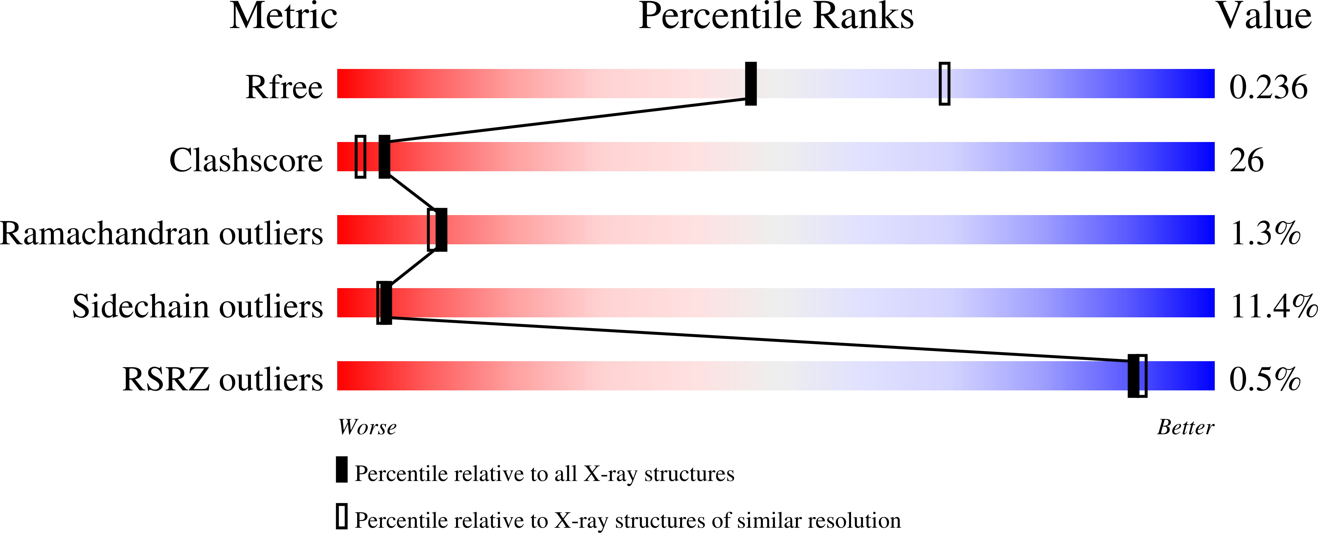
wwPDB Validation   3D Report Full Report


Ligand Structure Quality Assessment 


This is version 1.5 of the entry. See complete history


Literature

The structural mechanism of GTP stabilized oligomerization and catalytic activation of the Toxoplasma gondii uracil phosphoribosyltransferase.

Schumacher, M.A.Bashor, C.J.Song, M.H.Otsu, K.Zhu, S.Parry, R.J.Ullman, B.Brennan, R.G.

(2002) Proc Natl Acad Sci U S A 99: 78-83

  • DOI: https://doi.org/10.1073/pnas.012399599
  • Primary Citation of Related Structures:  
    1JLR, 1JLS

  • PubMed Abstract: 

    Uracil phosphoribosyltransferase (UPRT) is a member of a large family of salvage and biosynthetic enzymes, the phosphoribosyltransferases, and catalyzes the transfer of ribose 5-phosphate from alpha-d-5-phosphoribosyl-1-pyrophosphate (PRPP) to the N1 nitrogen of uracil. The UPRT from the opportunistic pathogen Toxoplasma gondii represents a promising target for rational drug design, because it can create intracellular, lethal nucleotides from subversive substrates. However, the development of such compounds requires a detailed understanding of the catalytic mechanism. Toward this end we determined the crystal structure of the T. gondii UPRT bound to uracil and cPRPP, a nonhydrolyzable PRPP analogue, to 2.5-A resolution. The structure suggests that the catalytic mechanism is substrate-assisted, and a tetramer would be the more active oligomeric form of the enzyme. Subsequent biochemical studies revealed that GTP binding, which has been suggested to play a role in catalysis by other UPRTs, causes a 6-fold activation of the T. gondii enzyme and strikingly stabilizes the tetramer form. The basis for stabilization was revealed in the 2.45-A resolution structure of the UPRT-GTP complex, whereby residues from three subunits contributed to GTP binding. Thus, our studies reveal an allosteric mechanism involving nucleotide stabilization of a more active, higher order oligomer. Such regulation of UPRT could play a role in the balance of purine and pyrimidine nucleotide pools in the cell.


  • Organizational Affiliation

    Department of Biochemistry and Molecular Biology, Oregon Health and Science University, 3181 Southwest Sam Jackson Park Road, Portland, OR 97201-3098, USA.


Macromolecules
Find similar proteins by:  (by identity cutoff)  |  3D Structure
Entity ID: 1
MoleculeChains Sequence LengthOrganismDetailsImage
Uracil PhosphoribosyltransferaseA,
B,
C [auth D],
D [auth C]
243Toxoplasma gondiiMutation(s): 1 
EC: 2.4.2.9
UniProt
Find proteins for Q26998 (Toxoplasma gondii)
Explore Q26998 
Go to UniProtKB:  Q26998
Entity Groups  
Sequence Clusters30% Identity50% Identity70% Identity90% Identity95% Identity100% Identity
UniProt GroupQ26998
Sequence Annotations
Expand
  • Reference Sequence
Small Molecules
Ligands 2 Unique
IDChains Name / Formula / InChI Key2D Diagram3D Interactions
GTP
Query on GTP

Download Ideal Coordinates CCD File 
G [auth A],
J [auth B],
M [auth D],
P [auth C]
GUANOSINE-5'-TRIPHOSPHATE
C10 H16 N5 O14 P3
XKMLYUALXHKNFT-UUOKFMHZSA-N
PO4
Query on PO4

Download Ideal Coordinates CCD File 
E [auth A]
F [auth A]
H [auth B]
I [auth B]
K [auth D]
E [auth A],
F [auth A],
H [auth B],
I [auth B],
K [auth D],
L [auth D],
N [auth C],
O [auth C]
PHOSPHATE ION
O4 P
NBIIXXVUZAFLBC-UHFFFAOYSA-K
Binding Affinity Annotations 
IDSourceBinding Affinity
GTP PDBBind:  1JLR Kd: 4.65e+5 (nM) from 1 assay(s)
Experimental Data & Validation

Experimental Data

  • Method: X-RAY DIFFRACTION
  • Resolution: 2.45 Å
  • R-Value Free: 0.242 
  • R-Value Work: 0.184 
  • Space Group: P 1 21 1
Unit Cell:
Length ( Å )Angle ( ˚ )
a = 60.6α = 90
b = 141.8β = 115
c = 71.4γ = 90
Software Package:
Software NamePurpose
CNSrefinement
bioteXdata reduction
bioteXdata scaling
CNSphasing

Structure Validation

View Full Validation Report


Ligand Structure Quality Assessment 


Entry History 

Deposition Data

Revision History  (Full details and data files)

  • Version 1.0: 2002-01-10
    Type: Initial release
  • Version 1.1: 2008-04-27
    Changes: Version format compliance
  • Version 1.2: 2011-07-13
    Changes: Version format compliance
  • Version 1.3: 2017-10-04
    Changes: Refinement description
  • Version 1.4: 2021-10-27
    Changes: Database references, Derived calculations
  • Version 1.5: 2023-08-16
    Changes: Data collection, Refinement description