Crystal Structure of the RuvA-RuvB Complex: A Structural Basis for the Holliday Junction Migrating Motor Machinery
Yamada, K., Miyata, T., Tsuchiya, D., Oyama, T., Fujiwara, Y., Ohnishi, T., Iwasaki, H., Shinagawa, H., Ariyoshi, M., Mayanagi, K., Morikawa, K.(2002) Mol Cell 10: 671-681
- PubMed: 12408833
- DOI: https://doi.org/10.1016/s1097-2765(02)00641-x
- Primary Citation of Related Structures:
1IXR, 1IXS - PubMed Abstract:
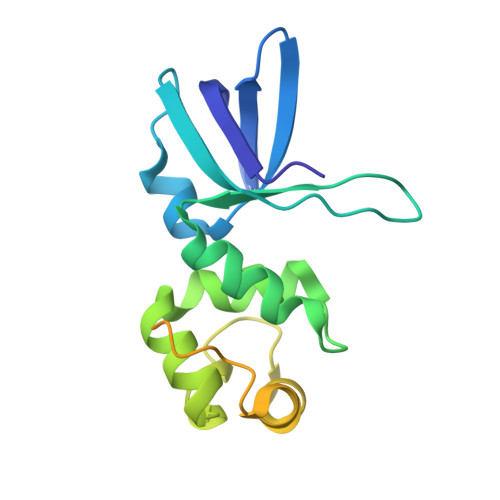
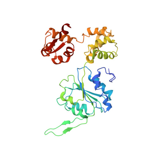
We present the X-ray structure of the RuvA-RuvB complex, which plays a crucial role in ATP-dependent branch migration. Two RuvA tetramers form the symmetric and closed octameric shell, where four RuvA domain IIIs spring out in the two opposite directions to be individually caught by a single RuvB. The binding of domain III deforms the protruding beta hairpin in the N-terminal domain of RuvB and thereby appears to induce a functional and less symmetric RuvB hexameric ring. The model of the RuvA-RuvB junction DNA ternary complex, constructed by fitting the X-ray structure into the averaged electron microscopic images of the RuvA-RuvB junction, appears to be more compatible with the branch migration mode of a fixed RuvA-RuvB interaction than with a rotational interaction mode.
Organizational Affiliation:
Biomolecular Engineering Research Institute, Suita, Osaka, Japan.


