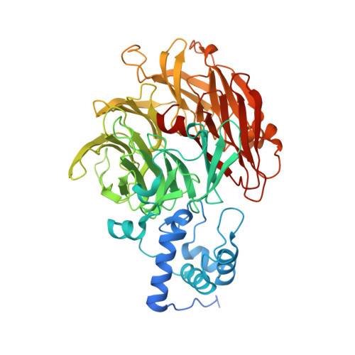
1H9X

Cytochrome cd1 Nitrite Reductase, reduced form


Experimental Data Snapshot

  • Method: X-RAY DIFFRACTION
  • Resolution: 2.10 Å
  • R-Value Free: 0.241 
  • R-Value Work: 0.216 

Starting Model: experimental
View more details

wwPDB Validation   3D Report Full Report


Ligand Structure Quality Assessment 


This is version 1.7 of the entry. See complete history


Literature

The Structure of an Alternative Form of Paracoccus Pantotrophus Cytochrome Cd1 Nitrite Reductase

Sjogren, T.Hajdu, J.

(2001) J Biol Chem 276: 29450

  • DOI: https://doi.org/10.1074/jbc.M103657200
  • Primary Citation of Related Structures:  
    1H9X, 1H9Y, 1HCM

  • PubMed Abstract: 

    Cytochrome cd(1) nitrite reductase is a bifunctional enzyme, which can catalyze the 1-electron reduction of nitrite to nitric oxide and the 4-electron reduction of dioxygen to water. Here we describe the structure of reduced nitrite reductase, crystallized under anaerobic conditions. The structure reveals substantial domain rearrangements with the c domain rotated by 60 degrees and shifted by approximately 20 A compared with previously known structures from crystals grown under oxidizing conditions. This alternative conformation gives rise to different electron transfer routes between the c and d(1) domains and points to the involvement of elements of very large structural changes in the function in this enzyme. In the present structure, the c heme has a His-69/Met-106 ligation, and this ligation does not change upon oxidation in the crystal. The d(1) heme is penta-coordinated, and the d(1) iron is displaced from the heme plane by 0.5 A toward the proximal ligand, His-200. After oxidation, the iron moves into the d(1) heme plane. A surprising finding is that although reduced nitrite reductase can be readily oxidized by dioxygen in the new crystal, it cannot turnover with its other substrate, nitrite. The results suggest that the rearrangement of the domains affects catalysis and substrate selectivity.


  • Organizational Affiliation

    Department of Biochemistry, Uppsala University, Box 576, S-751 23 Uppsala, Sweden.


Macromolecules
Find similar proteins by:  (by identity cutoff)  |  3D Structure
Entity ID: 1
MoleculeChains Sequence LengthOrganismDetailsImage
CYTOCHROME CD1 NITRITE REDUCTASE
A, B
567Paracoccus pantotrophusMutation(s): 0 
EC: 1.7.99.1 (UniProt), 1.7.2.1 (UniProt)
UniProt
Find proteins for P72181 (Paracoccus pantotrophus)
Explore P72181 
Go to UniProtKB:  P72181
Entity Groups  
Sequence Clusters30% Identity50% Identity70% Identity90% Identity95% Identity100% Identity
UniProt GroupP72181
Sequence Annotations
Expand
  • Reference Sequence
Small Molecules
Ligands 4 Unique
IDChains Name / Formula / InChI Key2D Diagram3D Interactions
DHE
Query on DHE

Download Ideal Coordinates CCD File 
D [auth A],
F [auth B]
HEME D
C34 H32 Fe N4 O10
XLQCGNUTSJTZNF-YDXXJHAFSA-L
HEC
Query on HEC

Download Ideal Coordinates CCD File 
C [auth A],
E [auth B]
HEME C
C34 H34 Fe N4 O4
HXQIYSLZKNYNMH-LJNAALQVSA-N
NHE
Query on NHE

Download Ideal Coordinates CCD File 
G [auth B]2-[N-CYCLOHEXYLAMINO]ETHANE SULFONIC ACID
C8 H17 N O3 S
MKWKNSIESPFAQN-UHFFFAOYSA-N
SO4
Query on SO4

Download Ideal Coordinates CCD File 
H [auth B],
I [auth B],
J [auth B]
SULFATE ION
O4 S
QAOWNCQODCNURD-UHFFFAOYSA-L
Experimental Data & Validation

Experimental Data

  • Method: X-RAY DIFFRACTION
  • Resolution: 2.10 Å
  • R-Value Free: 0.241 
  • R-Value Work: 0.216 
  • Space Group: P 43 21 2
Unit Cell:
Length ( Å )Angle ( ˚ )
a = 127.97α = 90
b = 127.97β = 90
c = 264.13γ = 90
Software Package:
Software NamePurpose
REFMACrefinement
DENZOdata reduction
SCALEPACKdata scaling

Structure Validation

View Full Validation Report


Ligand Structure Quality Assessment 


Entry History 

Deposition Data

Revision History  (Full details and data files)

  • Version 1.0: 2001-08-09
    Type: Initial release
  • Version 1.1: 2011-05-07
    Changes: Version format compliance
  • Version 1.2: 2011-07-13
    Changes: Version format compliance
  • Version 1.3: 2018-01-17
    Changes: Data collection
  • Version 1.4: 2019-05-08
    Changes: Data collection, Derived calculations, Experimental preparation
  • Version 1.5: 2020-03-04
    Changes: Derived calculations, Other
  • Version 1.6: 2023-12-13
    Changes: Data collection, Database references, Derived calculations, Refinement description
  • Version 1.7: 2024-10-23
    Changes: Structure summary