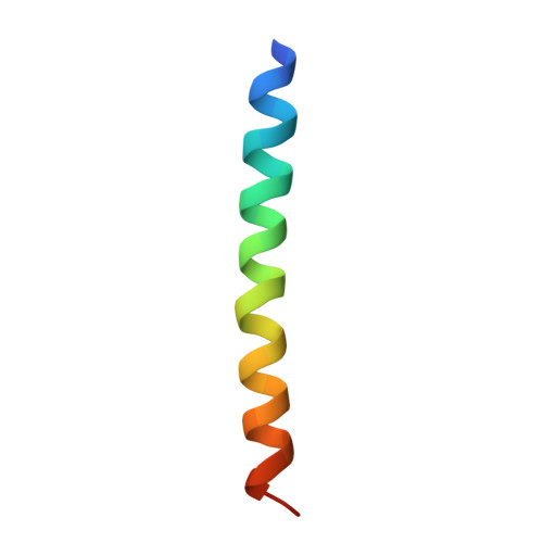
A switch between two-, three-, and four-stranded coiled coils in GCN4 leucine zipper mutants.
Harbury, P.B., Zhang, T., Kim, P.S., Alber, T.(1993) Science 262: 1401-1407
- PubMed: 8248779
- DOI: https://doi.org/10.1126/science.8248779
- Primary Citation of Related Structures:
1GCL - PubMed Abstract:
Coiled-coil sequences in proteins consist of heptad repeats containing two characteristic hydrophobic positions. The role of these buried hydrophobic residues in determining the structures of coiled coils was investigated by studying mutants of the GCN4 leucine zipper. When sets of buried residues were altered, two-, three-, and four-helix structures were formed. The x-ray crystal structure of the tetramer revealed a parallel, four-stranded coiled coil. In the tetramer conformation, the local packing geometry of the two hydrophobic positions in the heptad repeat is reversed relative to that in the dimer. These studies demonstrate that conserved, buried residues in the GCN4 leucine zipper direct dimer formation. In contrast to proposals that the pattern of hydrophobic and polar amino acids in a protein sequence is sufficient to determine three-dimensional structure, the shapes of buried side chains in coiled coils are essential determinants of the global fold.
Organizational Affiliation:
Department of Biological Chemistry and Molecular Pharmacology, Harvard Medical School, Boston, MA 02115.


