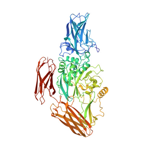
1FIE

RECOMBINANT HUMAN COAGULATION FACTOR XIII


Experimental Data Snapshot

  • Method: X-RAY DIFFRACTION
  • Resolution: 2.50 Å
  • R-Value Work: 0.182 
  • R-Value Observed: 0.182 

wwPDB Validation   3D Report Full Report


This is version 1.3 of the entry. See complete history


Literature

Structural evidence that the activation peptide is not released upon thrombin cleavage of factor XIII.

Yee, V.C.Pedersen, L.C.Bishop, P.D.Stenkamp, R.E.Teller, D.C.

(1995) Thromb Res 78: 389-397

  • DOI: https://doi.org/10.1016/0049-3848(95)00072-y
  • Primary Citation of Related Structures:  
    1FIE

  • PubMed Abstract: 

    The three-dimensional structure of the recombinant human factor XIII a2 dimer after cleavage by thrombin has been determined by X-ray crystallography. Factor XIII zymogen was treated with bovine alpha-thrombin in the presence of 3 mM CaCl2, and the cleaved protein was crystallized from Tris buffered at pH 6.5 using ethanol as the precipitating agent. Refinement of the molecular model of thrombin-cleaved factor XIII against diffraction data from 10.0 to 2.5 A resolution has been carried out to give a crystallographic R factor of 18.2%. The structure of thrombin-cleaved factor XIII is remarkably similar to that of the zymogen: there are no large conformational changes in the protein and the 37 residue amino terminus activation peptide remains associated with the rest of the molecule. This work shows that the activation peptide, upon thrombin cleavage, has the same conformation and occupies the same position with respect to the rest of the molecule as it does in the zymogen structure.


  • Organizational Affiliation

    Department of Biochemistry, University of Washington, Seattle 98195, USA.


Macromolecules
Find similar proteins by:  (by identity cutoff)  |  3D Structure
Entity ID: 1
MoleculeChains Sequence LengthOrganismDetailsImage
COAGULATION FACTOR XIII
A, B
731Homo sapiensMutation(s): 0 
EC: 2.3.2.13
UniProt & NIH Common Fund Data Resources
Find proteins for P00488 (Homo sapiens)
Explore P00488 
Go to UniProtKB:  P00488
PHAROS:  P00488
GTEx:  ENSG00000124491 
Entity Groups  
Sequence Clusters30% Identity50% Identity70% Identity90% Identity95% Identity100% Identity
UniProt GroupP00488
Sequence Annotations
Expand
  • Reference Sequence
Experimental Data & Validation

Experimental Data

  • Method: X-RAY DIFFRACTION
  • Resolution: 2.50 Å
  • R-Value Work: 0.182 
  • R-Value Observed: 0.182 
  • Space Group: P 1 21 1
Unit Cell:
Length ( Å )Angle ( ˚ )
a = 101.1α = 90
b = 73.13β = 106.4
c = 134.7γ = 90
Software Package:
Software NamePurpose
X-PLORmodel building
X-PLORrefinement
R-AXISdata reduction
R-AXISdata scaling
X-PLORphasing

Structure Validation

View Full Validation Report


Entry History 

Deposition Data

Revision History  (Full details and data files)

  • Version 1.0: 1997-02-12
    Type: Initial release
  • Version 1.1: 2008-03-24
    Changes: Version format compliance
  • Version 1.2: 2011-07-13
    Changes: Version format compliance
  • Version 1.3: 2024-02-07
    Changes: Data collection, Database references, Other