Structural studies of a baboon (Papio sp.) plasma protein inhibitor of cholesteryl ester transferase.
Buchko, G.W., Rozek, A., Kanda, P., Kennedy, M.A., Cushley, R.J.(2000) Protein Sci 9: 1548-1558
- PubMed: 10975576
- DOI: https://doi.org/10.1110/ps.9.8.1548
- Primary Citation of Related Structures:
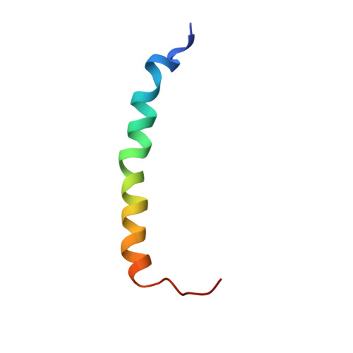
1EZE - PubMed Abstract:
A 38-residue protein associated with cholesteryl ester transfer inhibition has been identified in baboons (Papio sp.). The cholesteryl ester transfer inhibitor protein (CETIP) corresponds to the N-terminus of baboon apoC-I. Relative to CETIP, baboon apoC-I is a weak inhibitor of baboon cholesteryl ester transferase (CET). To study the structural features responsible for CET inhibition, CETIP was synthesized by solid-phase methods. Using sodium dodecyl sulfate (SDS) to model the lipoprotein environment, the solution structure of CETIP was probed by optical and 1H NMR spectroscopy. Circular dichroism data show that the protein lacks a well-defined structure in water but, upon the addition of SDS, becomes helical (56%). A small blue shift of 8 nm was observed in the intrinsic tryptophan fluorescence of CETIP in the presence of saturating amounts of SDS, suggesting that tryptophan-23 is not buried deeply in the lipid environment. The helical nature of CETIP in the presence of SDS was confirmed by upfield 1Halpha secondary shifts and an average solution structure determined by distance geometry/simulated annealing calculations using 476 NOE-based distance restraints. The backbone (N-Calpha-C=O) root-mean-square deviation of an ensemble of 17 out of 25 calculated structures superimposed on the average structure was 1.06+0.30 A using residues V4-P35 and 0.51+/-0.17 A using residues A7-S32. Although the side-chain orientations fit the basic description of a class A amphipathic helix, both intramolecular salt bridge formation and "snorkeling" of basic side chains toward the polar face play minor, if any, roles in stabilizing the lipid-bound amphipathic structure. Conformational features of the calculated structures for CETIP are discussed relative to models of CETIP inhibition of cholesteryl ester transferase.
Organizational Affiliation:
Institute of Molecular Biology and Biochemistry, Simon Fraser University, Burnaby, British Columbia, Canada. garry.buchko@pnl.gov