1EWY

ANABAENA PCC7119 FERREDOXIN:FERREDOXIN-NADP+-REDUCTASE COMPLEX


Experimental Data Snapshot

  • Method: X-RAY DIFFRACTION
  • Resolution: 2.38 Å
  • R-Value Free: 0.293 
  • R-Value Work: 0.219 
  • R-Value Observed: 0.232 

Starting Model: experimental
View more details

wwPDB Validation   3D Report Full Report


Ligand Structure Quality Assessment 


This is version 1.3 of the entry. See complete history


Literature

Crystallographic studies of the interaction between the ferredoxin-NADP+ reductase and ferredoxin from the cyanobacterium Anabaena: looking for the elusive ferredoxin molecule.

Morales, R.Kachalova, G.Vellieux, F.Charon, M.H.Frey, M.

(2000) Acta Crystallogr D Biol Crystallogr 56: 1408-1412

  • DOI: https://doi.org/10.1107/s0907444900010052
  • Primary Citation of Related Structures:  
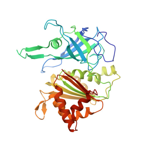
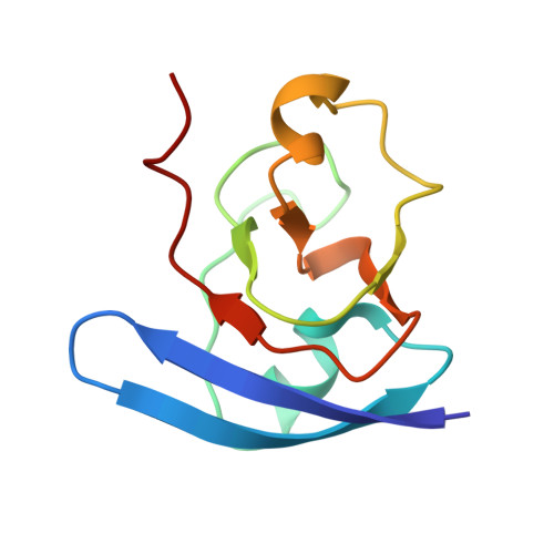
    1EWY

  • PubMed Abstract: 

    Ferredoxin-NADP(+) reductase (FNR) and its physiological electron donor ferredoxin (Fd) from the cyanobacterium Anabaena PCC7119 have been co-crystallized. The unit-cell parameters are a = b = 63.72, c = 158.02 A and the space group is P2(1)2(1)2(1). The crystal structure has been solved with 2.4 A resolution synchrotron data by molecular replacement, anomalous dispersion and R(min) search methods. For the computations, the crystal was treated as a merohedral twin. The asymmetric unit contains two FNR molecules and one ferredoxin molecule. The packing of the FNR molecules displays a nearly tetragonal symmetry (space group P4(3)2(1)2), whereas the ferredoxin arrangement is orthorhombic. This study provides the first crystallographic model of a dissociable complex between FNR and Fd.


  • Organizational Affiliation

    LCCP, Institut de Biologie Structurale J. P. Ebel, CEA-CNRS, 41 Rue Jules Horowitz, F38027 Grenoble, France.


Macromolecules
Find similar proteins by:  (by identity cutoff)  |  3D Structure
Entity ID: 1
MoleculeChains Sequence LengthOrganismDetailsImage
FERREDOXIN-NADP REDUCTASE
A, B
303Nostoc sp. PCC 7119Mutation(s): 0 
EC: 1.18.1.2
UniProt
Find proteins for P21890 (Nostoc sp. (strain ATCC 29151 / PCC 7119))
Explore P21890 
Go to UniProtKB:  P21890
Entity Groups  
Sequence Clusters30% Identity50% Identity70% Identity90% Identity95% Identity100% Identity
UniProt GroupP21890
Sequence Annotations
Expand
  • Reference Sequence
Find similar proteins by:  (by identity cutoff)  |  3D Structure
Entity ID: 2
MoleculeChains Sequence LengthOrganismDetailsImage
FERREDOXIN I98Nostoc sp. PCC 7119Mutation(s): 0 
UniProt
Find proteins for P0A3C8 (Nostoc sp. (strain ATCC 29151 / PCC 7119))
Explore P0A3C8 
Go to UniProtKB:  P0A3C8
Entity Groups  
Sequence Clusters30% Identity50% Identity70% Identity90% Identity95% Identity100% Identity
UniProt GroupP0A3C8
Sequence Annotations
Expand
  • Reference Sequence
Experimental Data & Validation

Experimental Data

  • Method: X-RAY DIFFRACTION
  • Resolution: 2.38 Å
  • R-Value Free: 0.293 
  • R-Value Work: 0.219 
  • R-Value Observed: 0.232 
  • Space Group: P 21 21 21
Unit Cell:
Length ( Å )Angle ( ˚ )
a = 63.73α = 90
b = 63.72β = 90
c = 158.02γ = 90
Software Package:
Software NamePurpose
AMoREphasing
SHELXL-97refinement
MOSFLMdata reduction
CCP4data scaling

Structure Validation

View Full Validation Report


Ligand Structure Quality Assessment 


Entry History 

Deposition Data

Revision History  (Full details and data files)

  • Version 1.0: 2001-02-07
    Type: Initial release
  • Version 1.1: 2008-04-27
    Changes: Version format compliance
  • Version 1.2: 2011-07-13
    Changes: Version format compliance
  • Version 1.3: 2023-08-09
    Changes: Data collection, Database references, Derived calculations, Refinement description