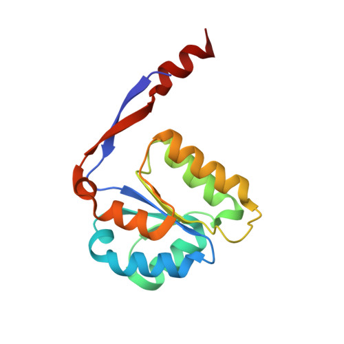
Mirroring perfection: the structure of methylglyoxal synthase complexed with the competitive inhibitor 2-phosphoglycolate.
Saadat, D., Harrison, D.H.(2000) Biochemistry 39: 2950-2960
- PubMed: 10715115
- DOI: https://doi.org/10.1021/bi992666f
- Primary Citation of Related Structures:
1EGH - PubMed Abstract:
The crystal structure of the transition-state analogue 2-phosphoglycolate (2PG) bound to methylglyoxal synthase (MGS) is presented at a resolution of 2.0 A. This structure is very similar to the previously determined structure of MGS complexed to formate and phosphate. Since 2PG is a competitive inhibitor of both MGS and triosephosphate isomerase (TIM), the carboxylate groups of each bound 2PG from this structure and the structure of 2PG bound to TIM were used to align and compare the active sites despite differences in their protein folds. The distances between the functional groups of Asp 71, His 98, His 19, and the carboxylate oxygens of the 2PG molecule in MGS are similar to the corresponding distances between the functional groups of Glu 165, His 95, Lys 13, and the carboxylate oxygens of the 2PG molecule in TIM. However, these spatial relationships are enantiomorphic to each other. Consistent with the known stereochemical data, the catalytic base Asp 71 is positioned on the opposite face of the 2PG-carboxylate plane as Glu 165 of TIM. Both His 98 of MGS and His 95 of TIM are in the plane of the carboxylate of 2PG, suggesting that these two residues are homologous in function. While His 19 of MGS and Lys 13 of TIM appear on the opposite face of the 2PG carboxylate plane, their relative location to the 2PG molecule is quite different, suggesting that they probably have different functions. Most remarkably, unlike the coplanar structure found in the 2PG molecule bound to TIM, the torsion angle around the C1-C2 bond of 2PG bound to MGS brings the phosphoryl moiety out of the molecule's carboxylate plane, facilitating elimination. Further, the superimposition of this structure with the structure of MGS bound to formate and phosphate suggests a model for the enzyme bound to the first transition state.
Organizational Affiliation:
Department of Biochemistry, Medical College of Wisconsin, 8701 Watertown Plank Road, Milwaukee, Wisconsin 53226, USA.


