Solution structure of murine epidermal growth factor determined by NMR spectroscopy and refined by energy minimization with restraints.
Montelione, G.T., Wuthrich, K., Burgess, A.W., Nice, E.C., Wagner, G., Gibson, K.D., Scheraga, H.A.(1992) Biochemistry 31: 236-249
- PubMed: 1731873
- DOI: https://doi.org/10.1021/bi00116a033
- Primary Citation of Related Structures:
1EGF, 3EGF - PubMed Abstract:
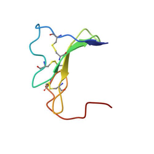
The solution structure of murine epidermal growth factor (mEGF) at pH 3.1 and a temperature of 28 degrees C has been determined from NMR data, using distance geometry calculations and restrained energy minimization. The structure determination is based on 730 conformational constraints derived from NMR data, including 644 NOE-derived upper bound distance constraints, constraints on the ranges of 32 dihedral angles based on measurements of vicinal coupling constants, and 54 upper and lower bound constraints associated with nine hydrogen bonds and the three disulfide bonds. The distance geometry interpretation of the NMR data is based on previously published sequence-specific 1H resonance assignments [Montelione et al. (1988) Biochemistry 27, 2235-2243], supplemented here with individual assignments for some side-chain amide, methylene, and isopropyl methyl protons. The molecular architecture of mEGF is the same as that described previously [Montelione et al. (1987) Proc. Natl. Acad. Sci. U.S.A. 84, 5226-5230], but the structure is overall more precisely determined by a more extensive set of NMR constraints. Analysis of proton NMR line widths, amide proton exchange rates, and side-chain 3J(H alpha-H beta) coupling constants provides evidence for internal motion in several regions of the mEGF molecule. Because mEGF is one member of a large family of homologous growth factors and protein domains for which X-ray crystal structures are not yet available, the atomic coordinates resulting from the present structure refinement (which we have deposited in the Brookhaven Protein Data Bank) are important data for understanding the structures of EGF-like proteins and for further detailed comparisons of these structures with mEGF.
Organizational Affiliation:
Center for Advanced Biotechnology and Medicine, Rutgers University, Piscataway, New Jersey 08854-5635.