Structural analysis of the heparin-binding site of the NC1 domain of collagen XIV by CD and NMR.
Montserret, R., Aubert-Foucher, E., McLeish, M.J., Hill, J.M., Ficheux, D., Jaquinod, M., van der Rest, M., Deleage, G., Penin, F.(1999) Biochemistry 38: 6479-6488
- PubMed: 10350466
- DOI: https://doi.org/10.1021/bi9900222
- Primary Citation of Related Structures:
1B9P, 1B9Q - PubMed Abstract:
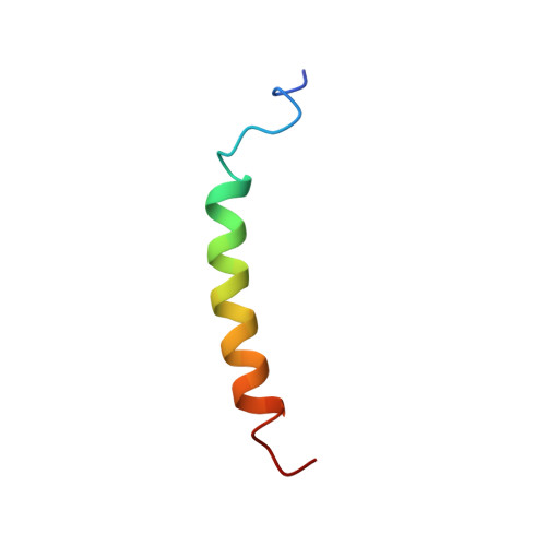
Type XIV collagen, a fibril-associated collagen with interrupted triple helices (FACIT), interacts with the surrounding extracellular matrix and/or with cells via its binding to glycosaminoglycans (GAGs). To further characterize such interactions in the NC1 domain of chicken collagen XIV, we identified amino acids essential for heparin binding by affinity chromatography analysis after proteolytic digestion of the synthetic peptide NC1(84-116). The 3D structure of this peptide was then obtained using circular dichroism and NMR. The NC1(84-116) peptide appeared poorly structured in water, but the stabilization of its conformation by the interaction with hydrophobic surfaces or by using cosolvents (TFE, SDS) revealed a high propensity to adopt an alpha-helical folding. A 3D structure model of NC1(84-116), calculated from NMR data recorded in a TFE/water mixture, showed that the NC1-heparin binding site forms a amphipathic alpha-helix exhibiting a twisted basic groove. It is structurally similar to the consensus spatial alpha-helix model of heparin-binding [Margalit et al. (1993) J. Biol. Chem. 268, 19228-19231], except that the GAG binding domain of NC1 may be extended over 18 residues, that is, the NC1(94-111) segment. In addition, the formation of a hydrophobic groove upon helix formation suggests the contribution of additional sequences to ensure the stability of the GAG-binding domain. Overall the NC1(84-116) model exhibits a nativelike conformation which presents suitably oriented residues for the interaction with a specific GAG.
Organizational Affiliation:
Institut de Biologie et de Chimie des Protéines, CNRS UPR 412, 7 passage du Vercors, 69367 Lyon Cedex 07, France.