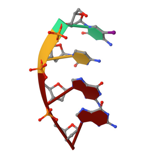
Helix geometry and hydration in an A-DNA tetramer: IC-C-G-G
Conner, B.N., Yoon, C., Dickerson, J.L., Dickerson, R.E.(1984) J Mol Biol 174: 663-695
- PubMed: 6726797
- DOI: https://doi.org/10.1016/0022-2836(84)90089-5
- Primary Citation of Related Structures:
1ANA - PubMed Abstract:
The DNA oligomer of sequence IC-C-G-G has been synthesized, and its X-ray crystal structure solved at a resolution of 2.0 A, using anomalous scattering from iodines in phase analysis: 48 cycles of Jack-Levitt restrained least-squares refinement resulted in a residual error of 19.9% over all data, or 16.5% for two-sigma data. Two double-helical tetramers stack in the crystal to form a continuous octamer, except for the two missing phosphate connections across the center. The octamer has a mean helix rotation of 33.7 degrees (10.7 base-pairs per turn), rise of 2.87 A, mean inclination angle of base-pairs of 14 degrees, and mean base-pair propeller twist of +16.3 degrees. Local variations in both helix rotation and base plane roll angles, including those across the center of the octamer, are as predicted from base sequence by sum functions sigma 1 and sigma 2. The three known DNA octamers: IC-C-G-G/IC-C-G-G, G-G-T-A-T-A-C-C and G-G-C-C-G-G-C-C, make up a graded series in this order, with monotonically changing structural parameters. An exhaustive comparison of torsion angle correlations among the known A helices confirms some structural expectations and reveals some new features. 86 water molecules have been located per double-helical IC-C-G-G tetramer (the asymmetric unit), of which 451/2 per tetramer lie within a first hydrogen-bonded shell of hydration. No ordered water structure is observed comparable to the minor groove spine of hydration in B-DNA.