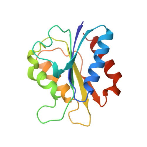
Modulation of the redox potentials of FMN in Desulfovibrio vulgaris flavodoxin: thermodynamic properties and crystal structures of glycine-61 mutants.
O'Farrell, P.A., Walsh, M.A., McCarthy, A.A., Higgins, T.M., Voordouw, G., Mayhew, S.G.(1998) Biochemistry 37: 8405-8416
- PubMed: 9622492
- DOI: https://doi.org/10.1021/bi973193k
- Primary Citation of Related Structures:
1AKR, 1AKT, 1AKW, 1AZL - PubMed Abstract:
Mutants of the electron-transfer protein flavodoxin from Desulfovibrio vulgaris were made by site-directed mutagenesis to investigate the role of glycine-61 in stabilizing the semiquinone of FMN by the protein and in controlling the flavin redox potentials. The spectroscopic properties, oxidation-reduction potentials, and flavin-binding properties of the mutant proteins, G61A/N/V and L, were compared with those of wild-type flavodoxin. The affinities of all of the mutant apoproteins for FMN and riboflavin were less than that of the wild-type apoprotein, and the redox potentials of the two 1-electron steps in the reduction of the complex with FMN were also affected by the mutations. Values for the dissociation constants of the complexes of the apoprotein with the semiquinone and hydroquinone forms of FMN were calculated from the redox potentials and the dissociation constant of the oxidized complex and used to derive the free energies of binding of the FMN in its three oxidation states. These showed that the semiquinone is destabilized in all of the mutants, and that the extent of destabilization tends to increase with increasing bulkiness of the side chain at residue 61. It is concluded that the hydrogen bond between the carbonyl of glycine-61 and N(5)H of FMN semiquinone in wild-type flavodoxin is either absent or severely impaired in the mutants. X-ray crystal structure analysis of the oxidized forms of the four mutant proteins shows that the protein loop that contains residue 61 is moved away from the flavin by 5-6 A. The hydrogen bond formed between the backbone nitrogen of aspartate-62 and O(4) of the dimethylisoalloxazine of the flavin in wild-type flavodoxin is absent in the mutants. Reliable structural information was not obtained for the reduced forms of the mutant proteins, but if the mutants change conformation when the flavin is reduced to the semiquinone, to facilitate hydrogen bonding between N(5)H and the carbonyl of residue 61, then the change must be different from that known to occur in wild-type flavodoxin.
Organizational Affiliation:
Department of Biochemistry, University College Dublin, Ireland.


