1AKE

STRUCTURE OF THE COMPLEX BETWEEN ADENYLATE KINASE FROM ESCHERICHIA COLI AND THE INHIBITOR AP5A REFINED AT 1.9 ANGSTROMS RESOLUTION: A MODEL FOR A CATALYTIC TRANSITION STATE


Experimental Data Snapshot

  • Method: X-RAY DIFFRACTION
  • Resolution: 2.00 Å
  • R-Value Work: 0.196 
  • R-Value Observed: 0.196 

wwPDB Validation   3D Report Full Report


Ligand Structure Quality Assessment 


This is version 1.2 of the entry. See complete history


Literature

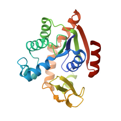
Structure of the complex between adenylate kinase from Escherichia coli and the inhibitor Ap5A refined at 1.9 A resolution. A model for a catalytic transition state.

Muller, C.W.Schulz, G.E.

(1992) J Mol Biol 224: 159-177

  • DOI: https://doi.org/10.1016/0022-2836(92)90582-5
  • Primary Citation of Related Structures:  
    1AKE

  • PubMed Abstract: 

    The structure of adenylate kinase from Escherichia coli ligated with the two-substrate-mimicking inhibitor P1,P5-bis(adenosine-5'-)pentaphosphate has been determined by X-ray diffraction and refined to a resolution of 1.9 A. The asymmetric unit of the crystals contains two copies of the complex, the structures of which agree well with each other. One of these copies is less well ordered in the crystals than the other, it shows generally higher temperature factors. The molecular packing in the crystals is discussed and correlated to crystal habit and anisotropic X-ray diffraction. The bound inhibitor simulates well the binding of substrates ATP and AMP, which are clearly assigned. The alpha-phosphate of AMP is well positioned for a nucleophilic attack on the gamma-phosphate of ATP. The observed structure readily allows the construction of a stabilized pentaco-ordinated transition state, as proposed for the known in-line mechanism of the enzyme, with nucleophile and leaving group in the apical positions of a trigonal bipyramid. The kinetic data of numerous mutations reported in the literature are correlated with the detailed structure of the enzyme. The mutants were classified. The concomitant increase of the Michaelis constants for ATP and AMP in the group of mutants that modify only the ATP-binding site cannot be explained.


  • Organizational Affiliation

    Institut für Organische Chemie und Biochemie der Universität, Freiburg, Germany.


Macromolecules
Find similar proteins by:  (by identity cutoff)  |  3D Structure
Entity ID: 1
MoleculeChains Sequence LengthOrganismDetailsImage
ADENYLATE KINASE
A, B
214Escherichia coliMutation(s): 0 
EC: 2.7.4.3
UniProt
Find proteins for P69441 (Escherichia coli (strain K12))
Explore P69441 
Go to UniProtKB:  P69441
Entity Groups  
Sequence Clusters30% Identity50% Identity70% Identity90% Identity95% Identity100% Identity
UniProt GroupP69441
Sequence Annotations
Expand
  • Reference Sequence
Small Molecules
Ligands 1 Unique
IDChains Name / Formula / InChI Key2D Diagram3D Interactions
AP5
Query on AP5

Download Ideal Coordinates CCD File 
C [auth A],
D [auth B]
BIS(ADENOSINE)-5'-PENTAPHOSPHATE
C20 H29 N10 O22 P5
OIMACDRJUANHTJ-XPWFQUROSA-N
Experimental Data & Validation

Experimental Data

  • Method: X-RAY DIFFRACTION
  • Resolution: 2.00 Å
  • R-Value Work: 0.196 
  • R-Value Observed: 0.196 
  • Space Group: P 21 2 21
Unit Cell:
Length ( Å )Angle ( ˚ )
a = 73.2α = 90
b = 79.8β = 90
c = 85γ = 90
Software Package:
Software NamePurpose
X-PLORmodel building
X-PLORrefinement
X-PLORphasing

Structure Validation

View Full Validation Report


Ligand Structure Quality Assessment 


Entry History 

Deposition Data

Revision History  (Full details and data files)

  • Version 1.0: 1994-01-31
    Type: Initial release
  • Version 1.1: 2008-03-24
    Changes: Version format compliance
  • Version 1.2: 2011-07-13
    Changes: Version format compliance