1A0I

ATP-DEPENDENT DNA LIGASE FROM BACTERIOPHAGE T7 COMPLEX WITH ATP


Experimental Data Snapshot

  • Method: X-RAY DIFFRACTION
  • Resolution: 2.60 Å
  • R-Value Free: 0.341 
  • R-Value Work: 0.222 
  • R-Value Observed: 0.222 

wwPDB Validation   3D Report Full Report


Ligand Structure Quality Assessment 


This is version 1.3 of the entry. See complete history


Literature

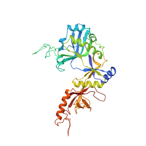
Crystal structure of an ATP-dependent DNA ligase from bacteriophage T7.

Subramanya, H.S.Doherty, A.J.Ashford, S.R.Wigley, D.B.

(1996) Cell 85: 607-615

  • DOI: https://doi.org/10.1016/s0092-8674(00)81260-x
  • Primary Citation of Related Structures:  
    1A0I

  • PubMed Abstract: 

    The crystal structure of the ATP-dependent DNA ligase from bacteriophage T7 has been solved at 2.6 A resolution. The protein comprises two domains with a deep cleft running between them. The structure of a complex with ATP reveals that the nucleotide binding pocket is situated on the larger N-terminal domain, at the base of the cleft between the two domains of the enzyme. Comparison of the overall domain structure with that of DNA methyltransferases, coupled with other evidence, suggests that DNA also binds in this cleft. Since this structure is the first of the nucleotidyltransferase superfamily, which includes the eukaryotic mRNA capping enzymes, the relationship between the structure of DNA ligase and that of other nucleotidyltransferases is also discussed.


  • Organizational Affiliation

    Laboratory of Molecular Biophysics, University of Oxford, United Kingdom.


Macromolecules
Find similar proteins by:  (by identity cutoff)  |  3D Structure
Entity ID: 1
MoleculeChains Sequence LengthOrganismDetailsImage
DNA LIGASE348Escherichia phage T7Mutation(s): 0 
Gene Names: LIG
EC: 6.5.1.1
UniProt
Find proteins for P00969 (Escherichia phage T7)
Explore P00969 
Go to UniProtKB:  P00969
Entity Groups  
Sequence Clusters30% Identity50% Identity70% Identity90% Identity95% Identity100% Identity
UniProt GroupP00969
Sequence Annotations
Expand
  • Reference Sequence
Small Molecules
Ligands 1 Unique
IDChains Name / Formula / InChI Key2D Diagram3D Interactions
ATP
Query on ATP

Download Ideal Coordinates CCD File 
B [auth A]ADENOSINE-5'-TRIPHOSPHATE
C10 H16 N5 O13 P3
ZKHQWZAMYRWXGA-KQYNXXCUSA-N
Experimental Data & Validation

Experimental Data

  • Method: X-RAY DIFFRACTION
  • Resolution: 2.60 Å
  • R-Value Free: 0.341 
  • R-Value Work: 0.222 
  • R-Value Observed: 0.222 
  • Space Group: P 21 21 2
Unit Cell:
Length ( Å )Angle ( ˚ )
a = 64.7α = 90
b = 85.2β = 90
c = 79.1γ = 90
Software Package:
Software NamePurpose
X-PLORmodel building
X-PLORrefinement
DENZOdata reduction
SCALEPACKdata scaling
X-PLORphasing

Structure Validation

View Full Validation Report


Ligand Structure Quality Assessment 


Entry History 

Deposition Data

Revision History  (Full details and data files)

  • Version 1.0: 1998-03-25
    Type: Initial release
  • Version 1.1: 2008-03-03
    Changes: Version format compliance
  • Version 1.2: 2011-07-13
    Changes: Version format compliance
  • Version 1.3: 2024-02-07
    Changes: Data collection, Database references, Derived calculations, Other