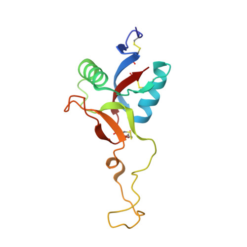
1X2W

Crystal Structure of Apo-Habu IX-bp at pH 4.6


Experimental Data Snapshot

  • Method: X-RAY DIFFRACTION
  • Resolution: 2.29 Å
  • R-Value Free: 0.280 
  • R-Value Work: 0.214 

Starting Model: experimental
View more details

wwPDB Validation   3D Report Full Report


This is version 1.5 of the entry. See complete history


Literature

pH-Dependent Structural Changes at Ca(2+)-binding sites of Coagulation Factor IX-binding Protein

Suzuki, N.Fujimoto, Z.Morita, T.Fukamizu, A.Mizuno, H.

(2005) J Mol Biol 353: 80-87

  • DOI: https://doi.org/10.1016/j.jmb.2005.08.018
  • Primary Citation of Related Structures:  
    1X2T, 1X2W

  • PubMed Abstract: 

    Coagulation factor IX-binding protein, isolated from Trimeresurus flavoviridis (IX-bp), is a C-type lectin-like protein. It is an anticoagulant consisting of homologous subunits, A and B. Each subunit has a Ca(2+)-binding site with a unique affinity (K(d) values of 14muM and 130muM at pH 7.5). These binding characteristics are pH-dependent and, under acidic conditions, the Ca(2+) binding of the low-affinity site was reduced considerably. In order to identify which site has high affinity and to investigate the pH-dependent Ca(2+) release mechanism, we have determined the crystal structures of IX-bp at pH 6.5 and pH 4.6 (apo form), and compared the Ca(2+)-binding sites with each other and with those of the solved structures under alkaline conditions; pH 7.8 and pH 8.0 (complexed form). At pH 6.5, Glu43 in the Ca(2+)-binding site of subunit A displayed two conformations. One (minor) is that in the alkaline state, and the other (major) is that at pH 4.6. However, the corresponding Gln43 residue of subunit B is in only a single conformation, which is almost identical with that in the alkaline state. At pH 4.6, Glu43 of subunit A adopts a conformation similar to that of the major conformer observed at pH 6.5, while Gln43 of subunit B assumes a new conformation, and both Ca(2+) positions are occupied by water molecules. These results showed that Glu43 of subunit A is much more sensitive to protonation than Gln43 of subunit B, and the conformational change of Glu43 occurs around pH6.5, which may correspond to the step of Ca(2+) release.


  • Organizational Affiliation

    Department of Biochemistry, National Institute of Agrobiological Sciences, Tsukuba, Ibaraki 305-8602, Japan.


Macromolecules
Find similar proteins by:  (by identity cutoff)  |  3D Structure
Entity ID: 1
MoleculeChains Sequence LengthOrganismDetailsImage
Coagulation factor IX/X-binding protein A chain129Protobothrops flavoviridisMutation(s): 0 
UniProt
Find proteins for Q7LZ71 (Protobothrops flavoviridis)
Explore Q7LZ71 
Go to UniProtKB:  Q7LZ71
Entity Groups  
Sequence Clusters30% Identity50% Identity70% Identity90% Identity95% Identity100% Identity
UniProt GroupQ7LZ71
Sequence Annotations
Expand
  • Reference Sequence
Find similar proteins by:  (by identity cutoff)  |  3D Structure
Entity ID: 2
MoleculeChains Sequence LengthOrganismDetailsImage
Coagulation factor IX/factor X-binding protein B chain123Protobothrops flavoviridisMutation(s): 0 
UniProt
Find proteins for P23807 (Protobothrops flavoviridis)
Explore P23807 
Go to UniProtKB:  P23807
Entity Groups  
Sequence Clusters30% Identity50% Identity70% Identity90% Identity95% Identity100% Identity
UniProt GroupP23807
Sequence Annotations
Expand
  • Reference Sequence
Experimental Data & Validation

Experimental Data

  • Method: X-RAY DIFFRACTION
  • Resolution: 2.29 Å
  • R-Value Free: 0.280 
  • R-Value Work: 0.214 
  • Space Group: C 1 2 1
Unit Cell:
Length ( Å )Angle ( ˚ )
a = 134.121α = 90
b = 37.831β = 110.46
c = 55.828γ = 90
Software Package:
Software NamePurpose
CNSrefinement
AMoREphasing

Structure Validation

View Full Validation Report


Entry History 

Deposition Data

Revision History  (Full details and data files)

  • Version 1.0: 2005-10-04
    Type: Initial release
  • Version 1.1: 2008-04-30
    Changes: Version format compliance
  • Version 1.2: 2011-07-13
    Changes: Version format compliance
  • Version 1.3: 2017-10-11
    Changes: Refinement description
  • Version 1.4: 2023-10-25
    Changes: Data collection, Database references, Derived calculations, Refinement description
  • Version 1.5: 2024-10-16
    Changes: Structure summary