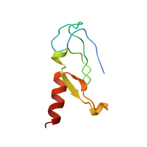
Crystal structure of a phosphatidylinositol 3-phosphate-specific membrane-targeting motif, the FYVE domain of Vps27p.
Misra, S., Hurley, J.H.(1999) Cell 97: 657-666
- PubMed: 10367894
- DOI: https://doi.org/10.1016/s0092-8674(00)80776-x
- Primary Citation of Related Structures:
1VFY - PubMed Abstract:
Phosphatidylinositol 3-phosphate regulates membrane trafficking and signaling pathways by interacting with the FYVE domains of target proteins. The 1.15 A structure of the Vps27p FYVE domain reveals two antiparallel beta sheets and an alpha helix stabilized by two Zn2+-binding clusters. The core secondary structures are similar to a rabphilin-3A Zn2+-binding domain and to the C1 and LIM domains. Phosphatidylinositol 3-phosphate binds to a pocket formed by the (R/K)(R/K)HHCR motif. A lattice contact shows how anionic ligands can interact with the phosphatidylinositol 3-phosphate-binding site. The tip of the FYVE domain has basic and hydrophobic surfaces positioned so that nonspecific interactions with the phospholipid bilayer can abet specific binding to phosphatidylinositol 3-phosphate.
Organizational Affiliation:
Laboratory of Molecular Biology, National Institute of Digestive, Diabetes, and Kidney Diseases, National Institutes of Health, Bethesda, Maryland 20892-0580, USA.