1SNA

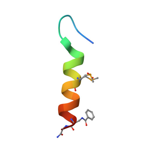
An Oligomeric Domain-Swapped Beta-Beta-Alpha Mini-Protein


Experimental Data Snapshot

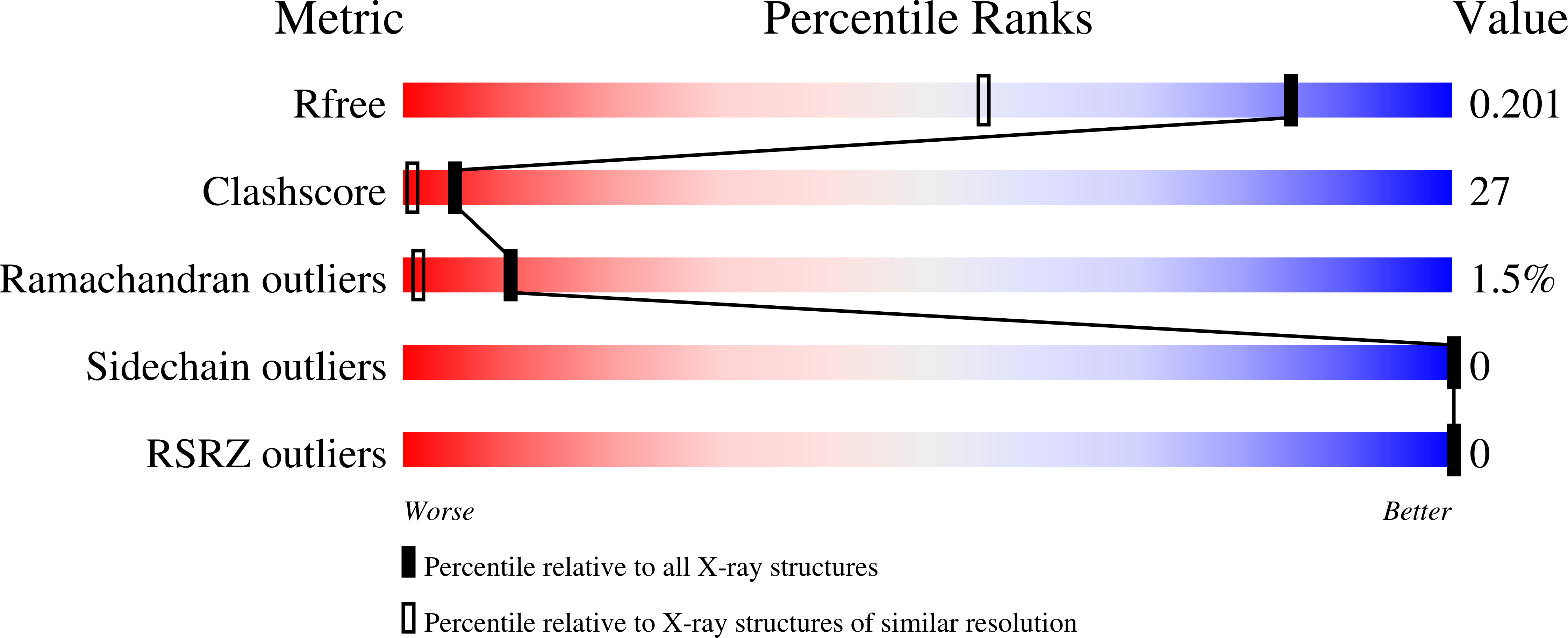
  • Method: X-RAY DIFFRACTION
  • Resolution: 1.50 Å
  • R-Value Free: 0.199 
  • R-Value Work: 0.161 
  • R-Value Observed: 0.161 

wwPDB Validation   3D Report Full Report


This is version 3.0 of the entry. See complete history


Literature

X-ray structure analysis of a designed oligomeric miniprotein reveals a discrete quaternary architecture.

Ali, M.H.Peisach, E.Allen, K.N.Imperiali, B.

(2004) Proc Natl Acad Sci U S A 101: 12183-12188

  • DOI: https://doi.org/10.1073/pnas.0401245101
  • Primary Citation of Related Structures:  
    1SN9, 1SNA, 1SNE

  • PubMed Abstract: 

    The x-ray crystal structure of an oligomeric miniprotein has been determined to a 1.2-A resolution by means of multiwavelength anomalous diffraction phasing with selenomethionine analogs that retain the biophysical characteristics of the native peptide. Peptide 1, comprising alpha and beta secondary structure elements with only 21 aa per monomer, associates as a discrete tetramer. The peptide adopts a previously uncharacterized quaternary structure in which alpha and beta components interact to form a tightly packed and well defined hydrophobic core. The structure provides insight into the origins of the unusual thermal stability of the oligomer. The miniprotein shares many characteristics of larger proteins, including cooperative folding, lack of 1-anilino-8-naphthalene sulfonate binding, and limited deuterium exchange, and possesses a buried surface area typical of native proteins.


  • Organizational Affiliation

    Department of Chemistry, Massachusetts Institute of Technology, 77 Massachusetts Avenue, Cambridge, MA 02139, USA.


Macromolecules

Find similar proteins by:  Sequence   |   3D Structure  

Entity ID: 1
MoleculeChains Sequence LengthOrganismDetailsImage
tetrameric beta-beta-alpha mini-protein
A, B, C, D
23N/AMutation(s): 0 
Entity Groups  
Sequence Clusters30% Identity50% Identity70% Identity90% Identity95% Identity100% Identity
Sequence Annotations
Expand
  • Reference Sequence
Small Molecules
Ligands 1 Unique
IDChains Name / Formula / InChI Key2D Diagram3D Interactions
IPA
Query on IPA

Download Ideal Coordinates CCD File 
E [auth C]ISOPROPYL ALCOHOL
C3 H8 O
KFZMGEQAYNKOFK-UHFFFAOYSA-N
Modified Residues  2 Unique
IDChains TypeFormula2D DiagramParent
DBZ
Query on DBZ
A, B, C, D
L-PEPTIDE LINKINGC10 H12 N2 O3ALA
MSO
Query on MSO
A, B, C, D
L-PEPTIDE LINKINGC5 H11 N O3 SeMET
Experimental Data & Validation

Experimental Data

  • Method: X-RAY DIFFRACTION
  • Resolution: 1.50 Å
  • R-Value Free: 0.199 
  • R-Value Work: 0.161 
  • R-Value Observed: 0.161 
  • Space Group: C 1 2 1
Unit Cell:
Length ( Å )Angle ( ˚ )
a = 50.027α = 90
b = 46.868β = 98.61
c = 31.426γ = 90
Software Package:
Software NamePurpose
CNSrefinement
DENZOdata reduction
SCALEPACKdata scaling
SOLVEphasing

Structure Validation

View Full Validation Report


Entry History 

Deposition Data

Revision History  (Full details and data files)

  • Version 1.0: 2004-08-17
    Type: Initial release
  • Version 1.1: 2008-04-29
    Changes: Version format compliance
  • Version 1.2: 2011-07-13
    Changes: Version format compliance
  • Version 1.3: 2011-11-16
    Changes: Atomic model
  • Version 2.0: 2023-11-15
    Changes: Atomic model, Data collection, Database references, Derived calculations
  • Version 3.0: 2024-05-08
    Changes: Atomic model