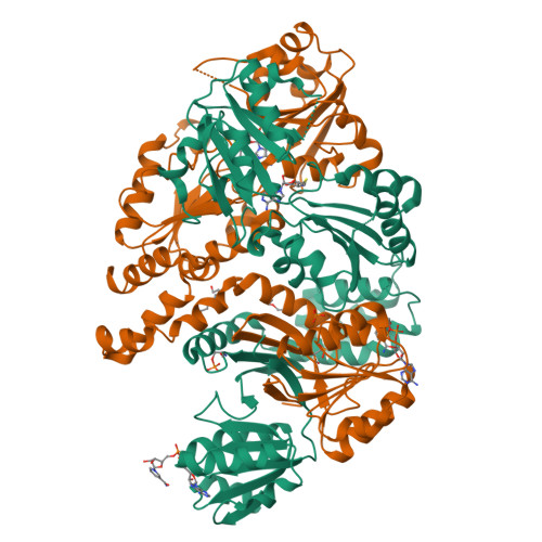
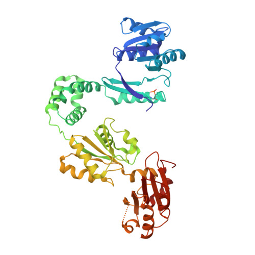
1PJS

The co-crystal structure of CysG, the multifunctional methyltransferase/dehydrogenase/ferrochelatase for siroheme synthesis, in complex with it NAD cofactor


Experimental Data Snapshot

  • Method: X-RAY DIFFRACTION
  • Resolution: 2.40 Å
  • R-Value Free: 0.281 
  • R-Value Work: 0.239 

Starting Model: experimental
View more details

wwPDB Validation   3D Report Full Report


Ligand Structure Quality Assessment 


This is version 1.5 of the entry. See complete history


Literature

CysG structure reveals tetrapyrrole-binding features and novel regulation of siroheme biosynthesis.

Stroupe, M.E.Leech, H.K.Daniels, D.S.Warren, M.J.Getzoff, E.D.

(2003) Nat Struct Biol 10: 1064-1073

  • DOI: https://doi.org/10.1038/nsb1007
  • Primary Citation of Related Structures:  
    1PJQ, 1PJS, 1PJT

  • PubMed Abstract: 

    Sulfur metabolism depends on the iron-containing porphinoid siroheme. In Salmonella enterica, the S-adenosyl-L-methionine (SAM)-dependent bismethyltransferase, dehydrogenase and ferrochelatase, CysG, synthesizes siroheme from uroporphyrinogen III (uro'gen III). The reactions mediated by CysG encompass two branchpoint intermediates in tetrapyrrole biosynthesis, diverting flux first from protoporphyrin IX biosynthesis and then from cobalamin (vitamin B(12)) biosynthesis. We determined the first structure of this multifunctional siroheme synthase by X-ray crystallography. CysG is a homodimeric gene fusion product containing two structurally independent modules: a bismethyltransferase and a dual-function dehydrogenase-chelatase. The methyltransferase active site is a deep groove with a hydrophobic patch surrounded by hydrogen bond donors. This asymmetric arrangement of amino acids may be important in directing substrate binding. Notably, our structure shows that CysG is a phosphoprotein. From mutational analysis of the post-translationally modified serine, we suggest a conserved role for phosphorylation in inhibiting dehydrogenase activity and modulating metabolic flux between siroheme and cobalamin pathways.


  • Organizational Affiliation

    Department of Molecular Biology and Skaggs Institute for Chemical Biology, The Scripps Research Institute, La Jolla, California 92037, USA.


Macromolecules
Find similar proteins by:  (by identity cutoff)  |  3D Structure
Entity ID: 1
MoleculeChains Sequence LengthOrganismDetailsImage
Siroheme synthase
A, B
457Salmonella enterica subsp. enterica serovar TyphimuriumMutation(s): 1 
Gene Names: CYSG OR STM3477
EC: 2.1.1.107 (PDB Primary Data), 1 (PDB Primary Data), 4.99.1 (PDB Primary Data), 4.99.1.4 (UniProt), 1.3.1.76 (UniProt)
UniProt
Find proteins for P25924 (Salmonella typhimurium (strain LT2 / SGSC1412 / ATCC 700720))
Explore P25924 
Go to UniProtKB:  P25924
Entity Groups  
Sequence Clusters30% Identity50% Identity70% Identity90% Identity95% Identity100% Identity
UniProt GroupP25924
Sequence Annotations
Expand
  • Reference Sequence
Small Molecules
Ligands 4 Unique
IDChains Name / Formula / InChI Key2D Diagram3D Interactions
NAD
Query on NAD

Download Ideal Coordinates CCD File 
D [auth A],
I [auth B]
NICOTINAMIDE-ADENINE-DINUCLEOTIDE
C21 H27 N7 O14 P2
BAWFJGJZGIEFAR-NNYOXOHSSA-N
SAH
Query on SAH

Download Ideal Coordinates CCD File 
C [auth A],
H [auth B]
S-ADENOSYL-L-HOMOCYSTEINE
C14 H20 N6 O5 S
ZJUKTBDSGOFHSH-WFMPWKQPSA-N
PGE
Query on PGE

Download Ideal Coordinates CCD File 
E [auth A],
F [auth A]
TRIETHYLENE GLYCOL
C6 H14 O4
ZIBGPFATKBEMQZ-UHFFFAOYSA-N
PO4
Query on PO4

Download Ideal Coordinates CCD File 
G [auth B]PHOSPHATE ION
O4 P
NBIIXXVUZAFLBC-UHFFFAOYSA-K
Modified Residues  1 Unique
IDChains TypeFormula2D DiagramParent
SEP
Query on SEP
A, B
L-PEPTIDE LINKINGC3 H8 N O6 PSER
Experimental Data & Validation

Experimental Data

  • Method: X-RAY DIFFRACTION
  • Resolution: 2.40 Å
  • R-Value Free: 0.281 
  • R-Value Work: 0.239 
  • Space Group: P 21 21 21
Unit Cell:
Length ( Å )Angle ( ˚ )
a = 60.124α = 90
b = 120.686β = 90
c = 130.178γ = 90
Software Package:
Software NamePurpose
CNSrefinement
DENZOdata reduction
SCALEPACKdata scaling
CNSphasing

Structure Validation

View Full Validation Report


Ligand Structure Quality Assessment 


Entry History 

Deposition Data

Revision History  (Full details and data files)

  • Version 1.0: 2003-12-02
    Type: Initial release
  • Version 1.1: 2008-04-29
    Changes: Version format compliance
  • Version 1.2: 2011-07-13
    Changes: Version format compliance
  • Version 1.3: 2014-11-12
    Changes: Structure summary
  • Version 1.4: 2023-08-16
    Changes: Data collection, Database references, Derived calculations, Refinement description
  • Version 1.5: 2024-11-06
    Changes: Data collection, Structure summary