1OZ3

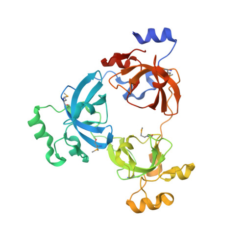
Crystal Structure of 3-MBT repeats of lethal (3) malignant Brain Tumor (Native-I) at 1.85 angstrom


Experimental Data Snapshot

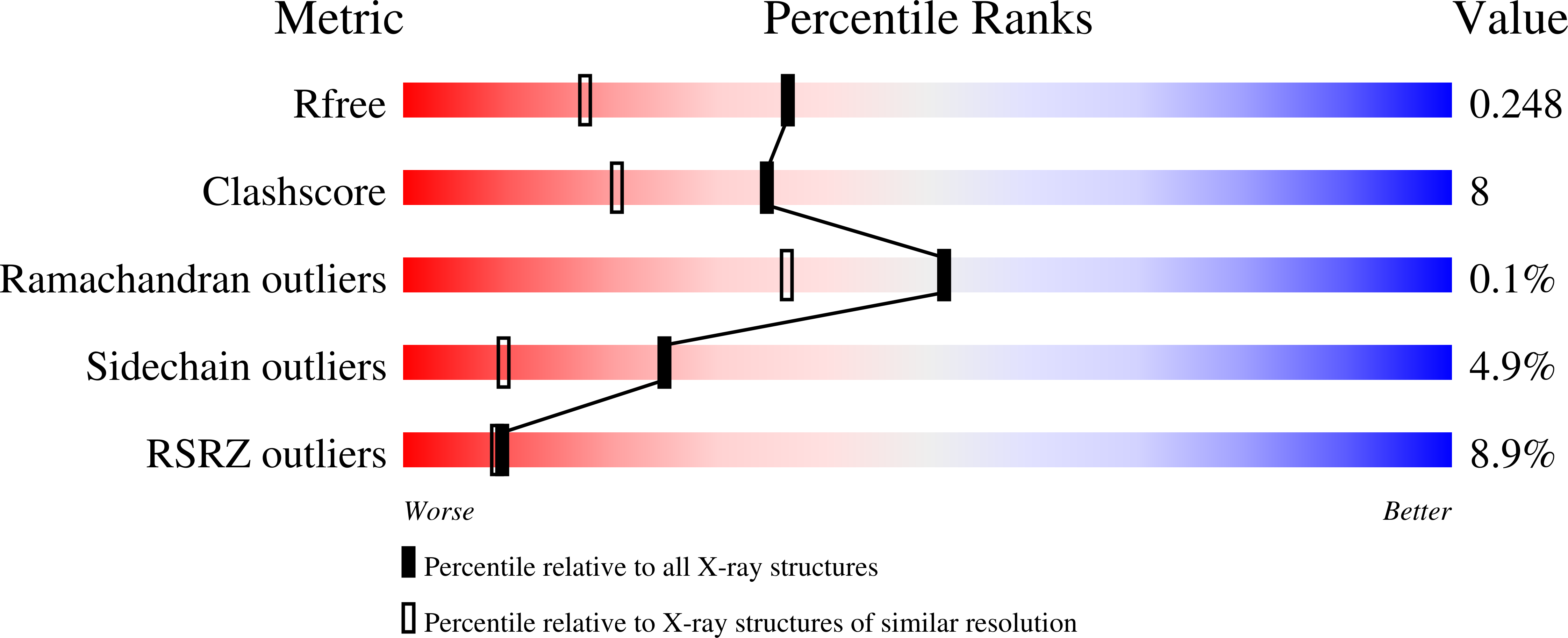
  • Method: X-RAY DIFFRACTION
  • Resolution: 1.85 Å
  • R-Value Free: 0.245 
  • R-Value Work: 0.211 
  • R-Value Observed: 0.213 

Starting Model: experimental
View more details

wwPDB Validation   3D Report Full Report


This is version 1.4 of the entry. See complete history


Literature

Malignant brain tumor repeats: a three-leaved propeller architecture with ligand/peptide binding pockets.

Wang, W.K.Tereshko, V.Boccuni, P.MacGrogan, D.Nimer, S.D.Patel, D.J.

(2003) Structure 11: 775-789

  • DOI: https://doi.org/10.1016/s0969-2126(03)00127-8
  • Primary Citation of Related Structures:  
    1OYX, 1OZ2, 1OZ3

  • PubMed Abstract: 

    We report on the X-ray structure of three 100-amino acid mbt repeats in h-l(3)mbt, a polycomb group protein involved in transcriptional repression, whose gene is located in a region of chromosome 20 associated with hematopoietic malignancies. Interdigitation between the extended arms and cores of the mbt repeats results in a three-leaved propeller-like architecture, containing a central cavity. We have identified one ligand binding pocket per mbt repeat, which accommodates either the morphilino ring of MES or the proline ring of the C-terminal peptide segment, within a cavity lined by aromatic amino acids. Strikingly, phenotypic alterations resulting from point mutations or deletions in the mbt repeats of the related Drosophila SCM protein are clustered in and around the ligand binding pocket.


  • Organizational Affiliation

    Cellular Biochemistry and Biophysics Program, Memorial Sloan-Kettering Cancer Center, New York, NY 10021, USA.


Macromolecules
Find similar proteins by:  (by identity cutoff)  |  3D Structure
Entity ID: 1
MoleculeChains Sequence LengthOrganismDetailsImage
Lethal(3)malignant brain tumor-like protein
A, B, C
331Homo sapiensMutation(s): 0 
Gene Names: L3MBTL OR L3MBT OR KIAA0681
UniProt & NIH Common Fund Data Resources
Find proteins for Q9Y468 (Homo sapiens)
Explore Q9Y468 
Go to UniProtKB:  Q9Y468
PHAROS:  Q9Y468
GTEx:  ENSG00000185513 
Entity Groups  
Sequence Clusters30% Identity50% Identity70% Identity90% Identity95% Identity100% Identity
UniProt GroupQ9Y468
Sequence Annotations
Expand
  • Reference Sequence
Small Molecules
Ligands 2 Unique
IDChains Name / Formula / InChI Key2D Diagram3D Interactions
MES
Query on MES

Download Ideal Coordinates CCD File 
F [auth A]
G [auth A]
M [auth B]
N [auth B]
R [auth C]
F [auth A],
G [auth A],
M [auth B],
N [auth B],
R [auth C],
S [auth C]
2-(N-MORPHOLINO)-ETHANESULFONIC ACID
C6 H13 N O4 S
SXGZJKUKBWWHRA-UHFFFAOYSA-N
SO4
Query on SO4

Download Ideal Coordinates CCD File 
D [auth A]
E [auth A]
H [auth B]
I [auth B]
J [auth B]
D [auth A],
E [auth A],
H [auth B],
I [auth B],
J [auth B],
K [auth B],
L [auth B],
O [auth C],
P [auth C],
Q [auth C]
SULFATE ION
O4 S
QAOWNCQODCNURD-UHFFFAOYSA-L
Modified Residues  1 Unique
IDChains TypeFormula2D DiagramParent
MSE
Query on MSE
A, B, C
L-PEPTIDE LINKINGC5 H11 N O2 SeMET
Experimental Data & Validation

Experimental Data

  • Method: X-RAY DIFFRACTION
  • Resolution: 1.85 Å
  • R-Value Free: 0.245 
  • R-Value Work: 0.211 
  • R-Value Observed: 0.213 
  • Space Group: P 32
Unit Cell:
Length ( Å )Angle ( ˚ )
a = 105.255α = 90
b = 105.255β = 90
c = 90.486γ = 120
Software Package:
Software NamePurpose
REFMACrefinement
HKL-2000data reduction
SCALEPACKdata scaling
AMoREphasing

Structure Validation

View Full Validation Report


Entry History 

Deposition Data

Revision History  (Full details and data files)

  • Version 1.0: 2003-08-19
    Type: Initial release
  • Version 1.1: 2008-04-29
    Changes: Version format compliance
  • Version 1.2: 2011-07-13
    Changes: Refinement description, Version format compliance
  • Version 1.3: 2023-08-16
    Changes: Data collection, Database references, Derived calculations, Refinement description
  • Version 1.4: 2023-11-15
    Changes: Data collection