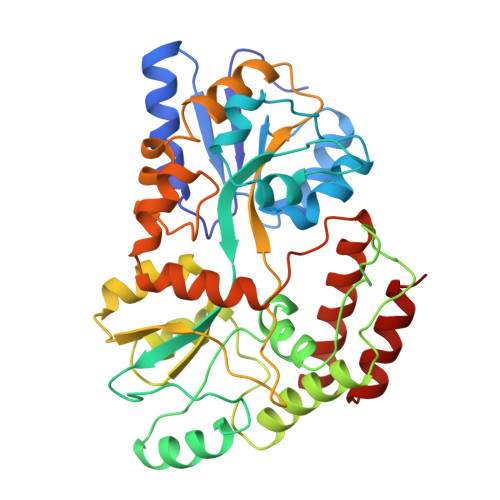
1MPD

MALTODEXTRIN-BINDING PROTEIN (MALTOSE-BINDING PROTEIN) MUTANT, WITH ARGININE REPLACING TRYPTOPHAN AT POSITION 230 (TRP-230-ARG), COMPLEXED WITH MALTOSE


Experimental Data Snapshot

  • Method: X-RAY DIFFRACTION
  • Resolution: 2.30 Å
  • R-Value Work: 0.168 
  • R-Value Observed: 0.168 

wwPDB Validation   3D Report Full Report


This is version 2.1 of the entry. See complete history


Literature

Crystal Structures and Solution Conformations of a Dominant-Negative Mutant of Escherichia Coli Maltose-Binding Protein

Shilton, B.H.Shuman, H.A.Mowbray, S.L.

(1996) J Mol Biol 264: 364-376

  • DOI: https://doi.org/10.1006/jmbi.1996.0646
  • Primary Citation of Related Structures:  
    1MPB, 1MPC, 1MPD

  • PubMed Abstract: 

    A mutant of the periplasmic maltose-binding protein (MBP) with altered transport properties was studied. A change of residue 230 from tryptophan to arginine results in dominant-negative MBP: expression of this protein against a wild-type background causes inhibition of maltose transport. As part of an investigation of the mechanism of such inhibition, we have solved crystal structures of both unliganded and liganded mutant protein. In the closed, liganded conformation, the side-chain of R230 projects into a region of the surface of MBP that has been identified as important for transport while in the open form, the same side-chain takes on a different, and less ordered, conformation. The crystallographic work is supplemented with a small-angle X-ray scattering study that provides evidence that the solution conformation of unliganded mutant is similar to that of wild-type MBP. It is concluded that dominant-negative inhibition of maltose transport must result from the formation of a non-productive complex between liganded-bound mutant MBP and wild-type MalFGK2. A general kinetic framework for transport by either wild-type MalFGK2 or MBP-independent MalFGK2 is used to understand the effects of dominant-negative MBP molecules on both of these systems.


  • Organizational Affiliation

    Department of Molecular Biology, Swedish Agricultural University, Uppsala, Sweden.


Macromolecules
Find similar proteins by:  (by identity cutoff)  |  3D Structure
Entity ID: 1
MoleculeChains Sequence LengthOrganismDetailsImage
MALTODEXTRIN-BINDING PROTEIN370Escherichia coli K-12Mutation(s): 0 
UniProt
Find proteins for P0AEX9 (Escherichia coli (strain K12))
Explore P0AEX9 
Go to UniProtKB:  P0AEX9
Entity Groups  
Sequence Clusters30% Identity50% Identity70% Identity90% Identity95% Identity100% Identity
UniProt GroupP0AEX9
Sequence Annotations
Expand
  • Reference Sequence
Oligosaccharides

Help

Entity ID: 2
MoleculeChains Length2D Diagram Glycosylation3D Interactions
alpha-D-glucopyranose-(1-4)-alpha-D-glucopyranose
B
2N/A
Glycosylation Resources
GlyTouCan:  G07411ON
GlyCosmos:  G07411ON
Biologically Interesting Molecules (External Reference) 1 Unique
Experimental Data & Validation

Experimental Data

  • Method: X-RAY DIFFRACTION
  • Resolution: 2.30 Å
  • R-Value Work: 0.168 
  • R-Value Observed: 0.168 
  • Space Group: P 21 21 21
Unit Cell:
Length ( Å )Angle ( ˚ )
a = 53.68α = 90
b = 58.96β = 90
c = 126.92γ = 90
Software Package:
Software NamePurpose
X-PLORmodel building
X-PLORrefinement
DENZOdata reduction
X-PLORphasing

Structure Validation

View Full Validation Report


Entry History 

Deposition Data

Revision History  (Full details and data files)

  • Version 1.0: 1995-10-15
    Type: Initial release
  • Version 1.1: 2008-03-24
    Changes: Version format compliance
  • Version 1.2: 2011-07-13
    Changes: Source and taxonomy, Version format compliance
  • Version 2.0: 2020-07-29
    Type: Remediation
    Reason: Carbohydrate remediation
    Changes: Atomic model, Data collection, Database references, Derived calculations, Non-polymer description, Other, Structure summary
  • Version 2.1: 2024-02-14
    Changes: Data collection, Database references, Structure summary