On the role of the conformational flexibility of the active-site lid on the allosteric kinetics of glucosamine-6-phosphate deaminase.
Bustos-Jaimes, I., Sosa-Peinado, A., Rudino-Pinera, E., Horjales, E., Calcagno, M.L.(2002) J Mol Biol 319: 183-189
- PubMed: 12051945
- DOI: https://doi.org/10.1016/S0022-2836(02)00096-7
- Primary Citation of Related Structures:
1JT9 - PubMed Abstract:
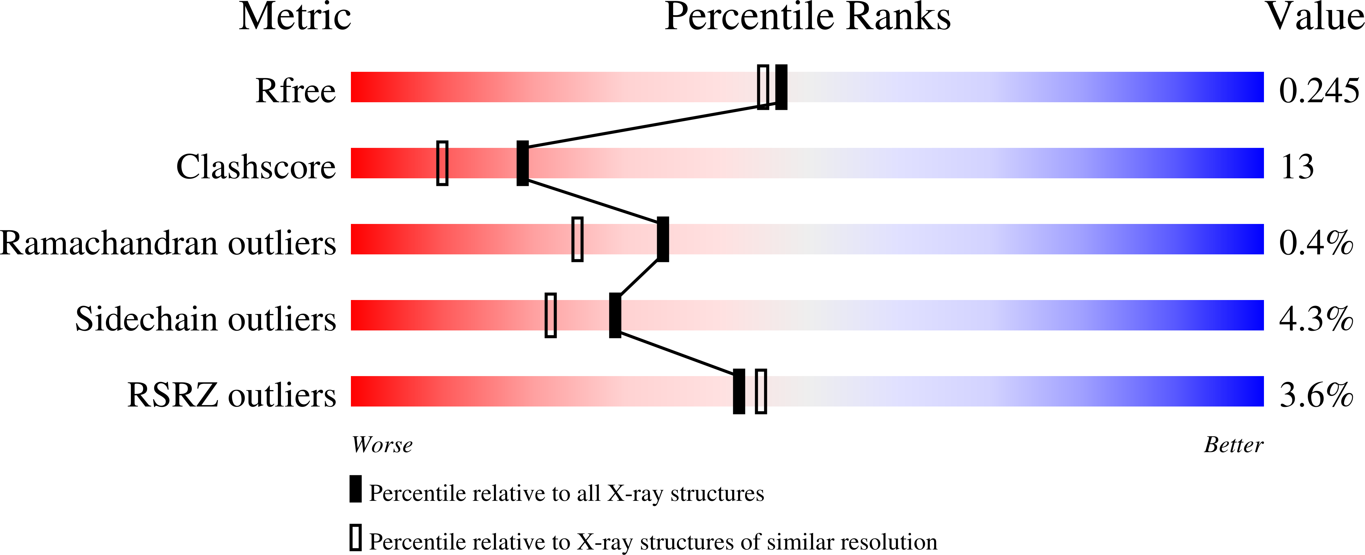
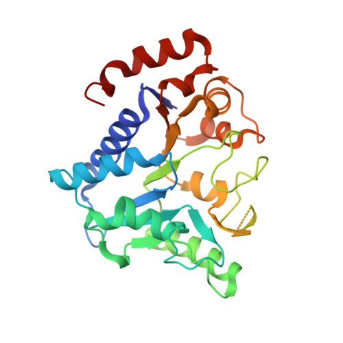
The active site of glucosamine-6-phosphate deaminase from Escherichia coli (GlcN6P deaminase, EC 3.5.99.6) has a complex lid formed by two antiparallel beta-strands connected by a helix-loop segment (158-187). This motif contains Arg172, which is a residue involved in binding the substrate in the active-site, and three residues that are part of the allosteric site, Arg158, Lys160 and Thr161. This dual binding role of the motif forming the lid suggests that it plays a key role in the functional coupling between active and allosteric sites. Previous crystallographic work showed that the temperature coefficients of the active-site lid are very large when the enzyme is in its T allosteric state. These coefficients decrease in the R state, thus suggesting that this motif changes its conformational flexibility as a consequence of the allosteric transition. In order to explore the possible connection between the conformational flexibility of the lid and the function of the deaminase, we constructed the site-directed mutant Phe174-Ala. Phe174 is located at the C-end of the lid helix and its side-chain establishes hydrophobic interactions with the remainder of the enzyme. The crystallographic structure of the T state of Phe174-Ala deaminase, determined at 2.02 A resolution, shows no density for the segment 162-181, which is part of the active-site lid (PDB 1JT9). This mutant form of the enzyme is essentially inactive in the absence of the allosteric activator, N-acetylglucosamine-6-P although it recovers its activity up to the wild-type level in the presence of this ligand. Spectrometric and binding studies show that inactivity is due to the inability of the active-site to bind ligands when the allosteric site is empty. These data indicate that the conformational flexibility of the active-site lid critically alters the binding properties of the active site, and that the occupation of the allosteric site restores the lid conformational flexibility to a functional state.
Organizational Affiliation:
Laboratorio de Fisicoquímica e Ingeniería de Proteínas, Departamento de Bioquímica, Facultad de Medicina, Universidad Nacional Autónoma de México, P.O. Box 70-159, Ciudad Universitaria, Mexico City, D.F. 04510, Mexico. ismaelb@servidor.unam.mx


