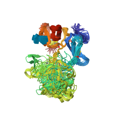
1E88

Solution structure of 6F11F22F2, a compact three-module fragment of the gelatin-binding domain of human fibronectin


Experimental Data Snapshot

  • Method: SOLUTION NMR
  • Conformers Calculated: 100 
  • Conformers Submitted: 20 
  • Selection Criteria: LEAST RESTRAINT VIOLATION 

wwPDB Validation   3D Report Full Report


This is version 1.5 of the entry. See complete history


Literature

The Hairpin Structure of the (6)F1(1)F2(2)F2 Fragment from Human Fibronectin Enhances Gelatin Binding

Pickford, A.R.Smith, S.P.Staunton, D.Boyd, J.Campbell, I.D.

(2001) EMBO J 20: 1519-1529

  • DOI: https://doi.org/10.1093/emboj/20.7.1519
  • Primary Citation of Related Structures:  
    1E88, 1E8B

  • PubMed Abstract: 

    The solution structure of the (6)F1(1)F2(2)F2 fragment from the gelatin-binding region of fibronectin has been determined (Protein Data Bank entry codes 1e88 and 1e8b). The structure reveals an extensive hydrophobic interface between the non-contiguous (6)F1 and (2)F2 modules. The buried surface area between (6)F1 and (2)F2 ( approximately 870 A(2)) is the largest intermodule interface seen in fibronectin to date. The dissection of (6)F1(1)F2(2)F2 into the (6)F1(1)F2 pair and (2)F2 results in near-complete loss of gelatin-binding activity. The hairpin topology of (6)F1(1)F2(2)F2 may facilitate intramolecular contact between the matrix assembly regions flanking the gelatin-binding domain. This is the first high-resolution study to reveal a compact, globular arrangement of modules in fibronectin. This arrangement is not consistent with the view that fibronectin is simply a linear 'string of beads'.


  • Organizational Affiliation

    Department of Biochemistry and Oxford Centre for Molecular Sciences, University of Oxford, South Parks Road, Oxford OX1 3QU, UK.


Macromolecules
Find similar proteins by:  (by identity cutoff)  |  3D Structure
Entity ID: 1
MoleculeChains Sequence LengthOrganismDetailsImage
FIBRONECTIN160Homo sapiensMutation(s): 0 
UniProt & NIH Common Fund Data Resources
Find proteins for P02751 (Homo sapiens)
Explore P02751 
Go to UniProtKB:  P02751
PHAROS:  P02751
GTEx:  ENSG00000115414 
Entity Groups  
Sequence Clusters30% Identity50% Identity70% Identity90% Identity95% Identity100% Identity
UniProt GroupP02751
Glycosylation
Glycosylation Sites: 1Go to GlyGen: P02751-15
Sequence Annotations
Expand
  • Reference Sequence
Small Molecules
Ligands 1 Unique
IDChains Name / Formula / InChI Key2D Diagram3D Interactions
NAG
Query on NAG

Download Ideal Coordinates CCD File 
B [auth A]2-acetamido-2-deoxy-beta-D-glucopyranose
C8 H15 N O6
OVRNDRQMDRJTHS-FMDGEEDCSA-N
Experimental Data & Validation

Experimental Data

  • Method: SOLUTION NMR
  • Conformers Calculated: 100 
  • Conformers Submitted: 20 
  • Selection Criteria: LEAST RESTRAINT VIOLATION 

Structure Validation

View Full Validation Report


Entry History 

Deposition Data

Revision History  (Full details and data files)

  • Version 1.0: 2000-10-09
    Type: Initial release
  • Version 1.1: 2011-10-19
    Changes: Non-polymer description, Other, Structure summary, Version format compliance
  • Version 1.2: 2012-11-28
    Changes: Database references, Derived calculations
  • Version 1.3: 2018-02-14
    Changes: Data collection, Database references, Source and taxonomy
  • Version 1.4: 2020-07-29
    Type: Remediation
    Reason: Carbohydrate remediation
    Changes: Advisory, Data collection, Derived calculations, Other, Structure summary
  • Version 1.5: 2024-10-16
    Changes: Data collection, Database references, Structure summary