1DQE

BOMBYX MORI PHEROMONE BINDING PROTEIN


Experimental Data Snapshot

  • Method: X-RAY DIFFRACTION
  • Resolution: 1.80 Å
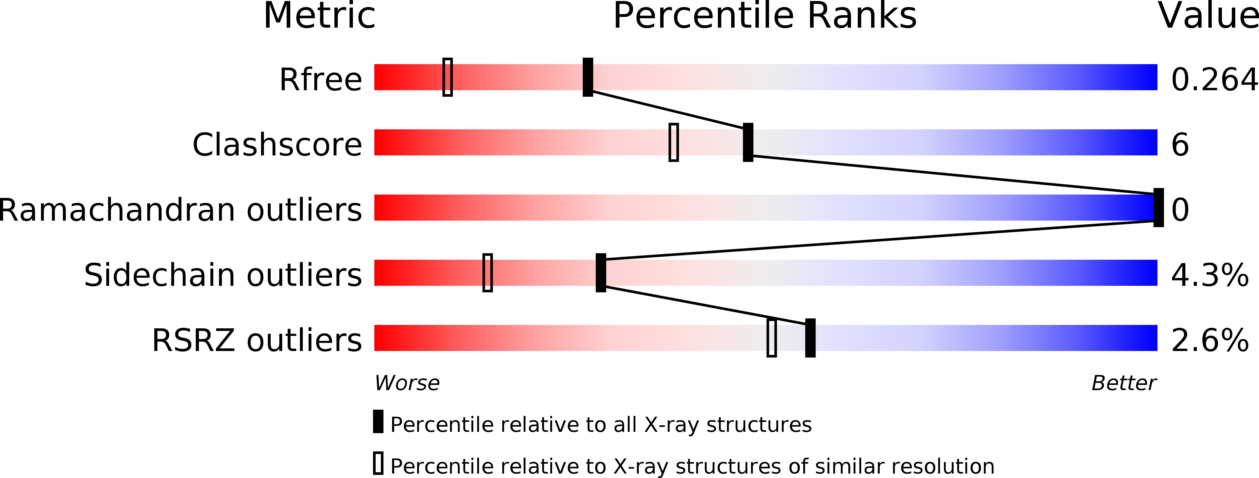
  • R-Value Free: 0.273 
  • R-Value Work: 0.210 

wwPDB Validation   3D Report Full Report


Ligand Structure Quality Assessment 


This is version 1.3 of the entry. See complete history


Literature

Sexual attraction in the silkworm moth: structure of the pheromone-binding-protein-bombykol complex.

Sandler, B.H.Nikonova, L.Leal, W.S.Clardy, J.

(2000) Chem Biol 7: 143-151

  • DOI: https://doi.org/10.1016/s1074-5521(00)00078-8
  • Primary Citation of Related Structures:  
    1DQE

  • PubMed Abstract: 

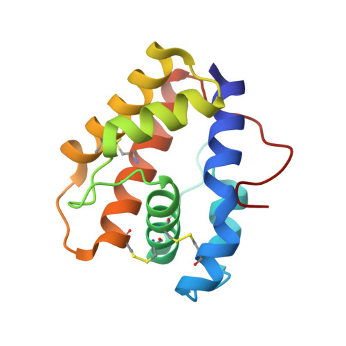
    Insects use volatile organic molecules to communicate messages with remarkable sensitivity and specificity. In one of the most studied systems, female silkworm moths (Bombyx mori) attract male mates with the pheromone bombykol, a volatile 16-carbon alcohol. In the male moth's antennae, a pheromone-binding protein conveys bombykol to a membrane-bound receptor on a nerve cell. The structure of the pheromone-binding protein, its binding and recognition of bombykol, and its full role in signal transduction are not known. The three-dimensional structure of the B. mori pheromone-binding protein with bound bombykol has been determined by X-ray diffraction at 1.8 A resolution. The pheromone binding protein of B. mori has six helices, and bombykol binds in a completely enclosed hydrophobic cavity formed by four antiparallel helices. Bombykol is bound in this cavity through numerous hydrophobic interactions, and sequence alignments suggest critical residues for specific pheromone binding.


  • Organizational Affiliation

    Department of Chemistry and Chemical Biology, Cornell University, Ithaca, NY 14853-1301, USA.


Macromolecules
Find similar proteins by:  (by identity cutoff)  |  3D Structure
Entity ID: 1
MoleculeChains Sequence LengthOrganismDetailsImage
PHEROMONE-BINDING PROTEIN
A, B
137Bombyx moriMutation(s): 0 
UniProt
Find proteins for P34174 (Bombyx mori)
Explore P34174 
Go to UniProtKB:  P34174
Entity Groups  
Sequence Clusters30% Identity50% Identity70% Identity90% Identity95% Identity100% Identity
UniProt GroupP34174
Sequence Annotations
Expand
  • Reference Sequence
Experimental Data & Validation

Experimental Data

  • Method: X-RAY DIFFRACTION
  • Resolution: 1.80 Å
  • R-Value Free: 0.273 
  • R-Value Work: 0.210 
  • Space Group: P 41 21 2
Unit Cell:
Length ( Å )Angle ( ˚ )
a = 53.18α = 90
b = 53.18β = 90
c = 191.54γ = 90
Software Package:
Software NamePurpose
MOSFLMdata reduction
SCALAdata scaling
SHARPphasing
REFMACrefinement
CCP4data scaling

Structure Validation

View Full Validation Report


Ligand Structure Quality Assessment 


Entry History 

Deposition Data

Revision History  (Full details and data files)

  • Version 1.0: 2001-01-10
    Type: Initial release
  • Version 1.1: 2008-04-27
    Changes: Version format compliance
  • Version 1.2: 2011-07-13
    Changes: Version format compliance
  • Version 1.3: 2024-10-16
    Changes: Data collection, Database references, Derived calculations, Structure summary