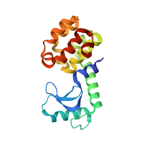
Methionine and alanine substitutions show that the formation of wild-type-like structure in the carboxy-terminal domain of T4 lysozyme is a rate-limiting step in folding.
Gassner, N.C., Baase, W.A., Lindstrom, J.D., Lu, J., Dahlquist, F.W., Matthews, B.W.(1999) Biochemistry 38: 14451-14460
- PubMed: 10545167
- DOI: https://doi.org/10.1021/bi9915519
- Primary Citation of Related Structures:
1CTW, 1CU0, 1CU2, 1CU3, 1CU5, 1CU6, 1CUP, 1CUQ, 1CV0, 1CV1, 1CV3, 1CV4, 1CV5, 1CV6, 1CVK, 1QSQ - PubMed Abstract:
In an attempt to identify a systematic relation between the structure of a protein and its folding kinetics, the rate of folding was determined for 20 mutants of T4 lysozyme in which a bulky, buried, nonpolar wild-type residue (Leu, Ile, Phe, Val, or Met) was substituted with alanine. Methionine, which approximated the size of the original side chain but which is of different shape and flexibility, was also substituted at most of the same sites. Mutations that substantially destabilize the protein and are located in the carboxy-terminal domain generally slow the rate of folding. Destabilizing mutations in the amino-terminal domain, however, have little effect on the rate of folding. Mutations that have little effect on stability tend to have little effect on the rate, no matter where they are located. These results suggest that, at the rate-limiting step, elements of structure in the C-terminal domain are formed and have a structure similar to that of the fully folded protein. Consistent with this, two variants that somewhat increase the rate of folding (Phe104 --> Met and Val149 --> Met) are located within the carboxy-terminal domain and maintain or improve packing with very little perturbation of the wild-type structure.
Organizational Affiliation:
Institute of Molecular Biology, Howard Hughes Medical Institute, and Departments of Chemistry and Physics, 1229 University of Oregon, Eugene, Oregon 97403-1229, USA.


