1A81

CRYSTAL STRUCTURE OF THE TANDEM SH2 DOMAIN OF THE SYK KINASE BOUND TO A DUALLY TYROSINE-PHOSPHORYLATED ITAM


Experimental Data Snapshot

  • Method: X-RAY DIFFRACTION
  • Resolution: 3.00 Å
  • R-Value Free: 0.317 
  • R-Value Work: 0.226 
  • R-Value Observed: 0.226 

wwPDB Validation   3D Report Full Report


This is version 1.3 of the entry. See complete history


Literature

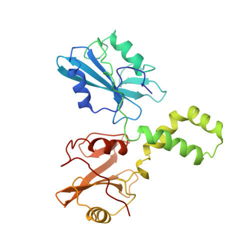
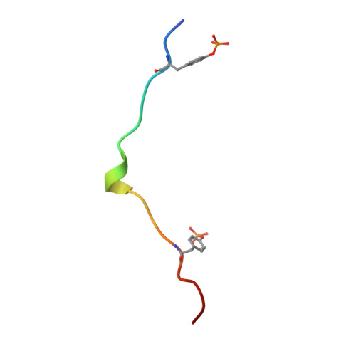
Structural basis for Syk tyrosine kinase ubiquity in signal transduction pathways revealed by the crystal structure of its regulatory SH2 domains bound to a dually phosphorylated ITAM peptide.

Futterer, K.Wong, J.Grucza, R.A.Chan, A.C.Waksman, G.

(1998) J Mol Biol 281: 523-537

  • DOI: https://doi.org/10.1006/jmbi.1998.1964
  • Primary Citation of Related Structures:  
    1A81

  • PubMed Abstract: 

    The Syk family of kinases, consisting of ZAP-70 and Syk, play essential roles in a variety of immune and non-immune cells. This family of kinases is characterized by the presence of two adjacent SH2 domains which mediate their localization to the membrane through receptor encoded tyrosine phosphorylated motifs. While these two kinases share many structural and functional features, the more ubiquitous nature of Syk has suggested that this kinase may accommodate a greater variety of motifs to mediate its function. We present the crystal structure of the tandem SH2 domain of Syk complexed with a dually phosphorylated ITAM peptide. The structure was solved by multiple isomorphous replacement at 3.0 A resolution. The asymmetric unit comprises six copies of the liganded protein, revealing a surprising flexibility in the relative orientation of the two SH2 domains. The C-terminal phosphotyrosine-binding site is very different from the equivalent region of ZAP-70, suggesting that in contrast to ZAP-70, the two SH2 domains of Syk can function as independent units. The conformational flexibility and structural independence of the SH2 modules of Syk likely provides the molecular basis for the more ubiquitous involvement of Syk in a variety of signal transduction pathways.


  • Organizational Affiliation

    Department of Biochemistry and Molecular Biophysics, Washington University School of Medicine, 660 South Euclid Av., Saint Louis, MO, 63110, USA.


Macromolecules
Find similar proteins by:  (by identity cutoff)  |  3D Structure
Entity ID: 1
MoleculeChains Sequence LengthOrganismDetailsImage
SYK KINASE
A, C, E, G, I
A, C, E, G, I, K
254Homo sapiensMutation(s): 0 
Gene Names: SYK
EC: 2.7.1.112
UniProt & NIH Common Fund Data Resources
Find proteins for P43405 (Homo sapiens)
Explore P43405 
Go to UniProtKB:  P43405
PHAROS:  P43405
GTEx:  ENSG00000165025 
Entity Groups  
Sequence Clusters30% Identity50% Identity70% Identity90% Identity95% Identity100% Identity
UniProt GroupP43405
Sequence Annotations
Expand
  • Reference Sequence

Find similar proteins by:  Sequence   |   3D Structure  

Entity ID: 2
MoleculeChains Sequence LengthOrganismDetailsImage
T-CELL SURFACE GLYCOPROTEIN CD3 EPSILON CHAIN
B, D, F, H, J
B, D, F, H, J, L
18Homo sapiensMutation(s): 2 
UniProt & NIH Common Fund Data Resources
Find proteins for P07766 (Homo sapiens)
Explore P07766 
Go to UniProtKB:  P07766
PHAROS:  P07766
GTEx:  ENSG00000198851 
Entity Groups  
Sequence Clusters30% Identity50% Identity70% Identity90% Identity95% Identity100% Identity
UniProt GroupP07766
Sequence Annotations
Expand
  • Reference Sequence
Small Molecules
Modified Residues  1 Unique
IDChains TypeFormula2D DiagramParent
PTR
Query on PTR
B, D, F, H, J
B, D, F, H, J, L
L-PEPTIDE LINKINGC9 H12 N O6 PTYR
Experimental Data & Validation

Experimental Data

  • Method: X-RAY DIFFRACTION
  • Resolution: 3.00 Å
  • R-Value Free: 0.317 
  • R-Value Work: 0.226 
  • R-Value Observed: 0.226 
  • Space Group: P 1 21 1
Unit Cell:
Length ( Å )Angle ( ˚ )
a = 85.5α = 90
b = 146.9β = 97.6
c = 91.5γ = 90
Software Package:
Software NamePurpose
SHARPphasing
X-PLORmodel building
X-PLORrefinement
DENZOdata reduction
SCALEPACKdata scaling
X-PLORphasing

Structure Validation

View Full Validation Report


Entry History 

Deposition Data

Revision History  (Full details and data files)

  • Version 1.0: 1998-10-21
    Type: Initial release
  • Version 1.1: 2008-03-24
    Changes: Version format compliance
  • Version 1.2: 2011-07-13
    Changes: Derived calculations, Version format compliance
  • Version 1.3: 2016-05-25
    Changes: Structure summary