7NCF

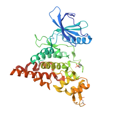
Crystal structure of HIPK2 in complex with MU135 (compound 21e)


Experimental Data Snapshot

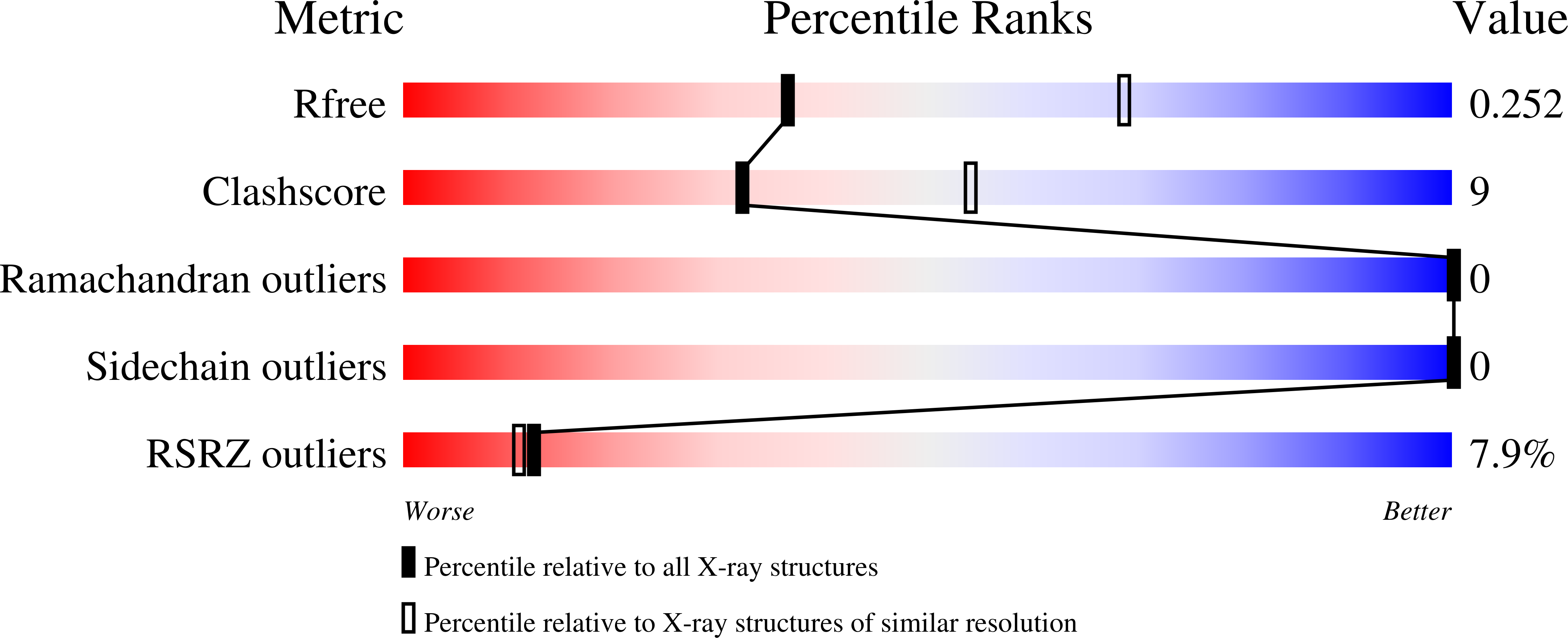
  • Method: X-RAY DIFFRACTION
  • Resolution: 2.72 Å
  • R-Value Free: 0.251 
  • R-Value Work: 0.195 
  • R-Value Observed: 0.197 

Starting Model: experimental
View more details

wwPDB Validation   3D Report Full Report


Ligand Structure Quality Assessment 


This is version 1.2 of the entry. See complete history


Literature

Highly selective inhibitors of protein kinases CLK and HIPK with the furo[3,2-b]pyridine core.

Nemec, V.Maier, L.Berger, B.T.Chaikuad, A.Drapela, S.Soucek, K.Knapp, S.Paruch, K.

(2021) Eur J Med Chem 215: 113299-113299

  • DOI: https://doi.org/10.1016/j.ejmech.2021.113299
  • Primary Citation of Related Structures:  
    7NCF

  • PubMed Abstract: 

    The furo [3,2-b]pyridine motif represents a relatively underexplored central pharmacophore in the area of kinase inhibitors. Herein, we report flexible synthesis of 3,5-disubstituted furo [3,2-b]pyridines that relies on chemoselective couplings of newly prepared 5-chloro-3-iodofuro [3,2-b]pyridine. This methodology allowed efficient second-generation synthesis of the state-of-the-art chemical biology probe for CLK1/2/4 MU1210, and identification of the highly selective inhibitors of HIPKs MU135 and MU1787 which are presented and characterized in this study, including the X-ray crystal structure of MU135 in HIPK2. chemical biology probe.


  • Organizational Affiliation

    Department of Chemistry, Faculty of Science, Masaryk University, Kamenice 5, 625 00, Brno, Czech Republic; International Clinical Research Center, Center for Biomolecular and Cellular Engineering, St. Anne's University Hospital in Brno, Pekařská 53, 656 91, Brno, Czech Republic.


Macromolecules
Find similar proteins by:  (by identity cutoff)  |  3D Structure
Entity ID: 1
MoleculeChains Sequence LengthOrganismDetailsImage
Homeodomain-interacting protein kinase 2367Homo sapiensMutation(s): 0 
Gene Names: HIPK2
EC: 2.7.11.1
UniProt & NIH Common Fund Data Resources
Find proteins for Q9H2X6 (Homo sapiens)
Explore Q9H2X6 
Go to UniProtKB:  Q9H2X6
PHAROS:  Q9H2X6
GTEx:  ENSG00000064393 
Entity Groups  
Sequence Clusters30% Identity50% Identity70% Identity90% Identity95% Identity100% Identity
UniProt GroupQ9H2X6
Sequence Annotations
Expand
  • Reference Sequence
Small Molecules
Ligands 1 Unique
IDChains Name / Formula / InChI Key2D Diagram3D Interactions
U82 (Subject of Investigation/LOI)
Query on U82

Download Ideal Coordinates CCD File 
B [auth A]3-(4-Tert-butylphenyl)-5-(1H-pyrazol-4-yl)furo[3,2-b]pyridine
C20 H19 N3 O
OOLXXVMFVSTNLJ-UHFFFAOYSA-N
Modified Residues  2 Unique
IDChains TypeFormula2D DiagramParent
PTR
Query on PTR
A
L-PEPTIDE LINKINGC9 H12 N O6 PTYR
SEP
Query on SEP
A
L-PEPTIDE LINKINGC3 H8 N O6 PSER
Binding Affinity Annotations 
IDSourceBinding Affinity
U82 BindingDB:  7NCF IC50: min: 119, max: 476 (nM) from 3 assay(s)
Experimental Data & Validation

Experimental Data

  • Method: X-RAY DIFFRACTION
  • Resolution: 2.72 Å
  • R-Value Free: 0.251 
  • R-Value Work: 0.195 
  • R-Value Observed: 0.197 
  • Space Group: P 62
Unit Cell:
Length ( Å )Angle ( ˚ )
a = 133.855α = 90
b = 133.855β = 90
c = 56.771γ = 120
Software Package:
Software NamePurpose
Aimlessdata scaling
REFMACrefinement
PDB_EXTRACTdata extraction
XDSdata reduction
PHASERphasing

Structure Validation

View Full Validation Report


Ligand Structure Quality Assessment 


Entry History 

Deposition Data

Revision History  (Full details and data files)

  • Version 1.0: 2021-03-03
    Type: Initial release
  • Version 1.1: 2021-04-14
    Changes: Database references
  • Version 1.2: 2024-01-31
    Changes: Data collection, Database references, Refinement description