7KAC

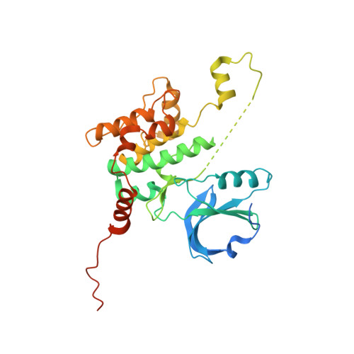
Crystal structure of HPK1 (MAP4K1) kinase in complex with 5-{[4-{[(1S)-2-HYDROXY-1-PHENYLETHYL]AMINO}-5-(1,3,4-OXADIAZOL-2-YL)PYRIMIDIN-2-YL]AMINO}-3,3-DIMETHYL-2-BENZOFURAN-1(3H)-ONE


Experimental Data Snapshot

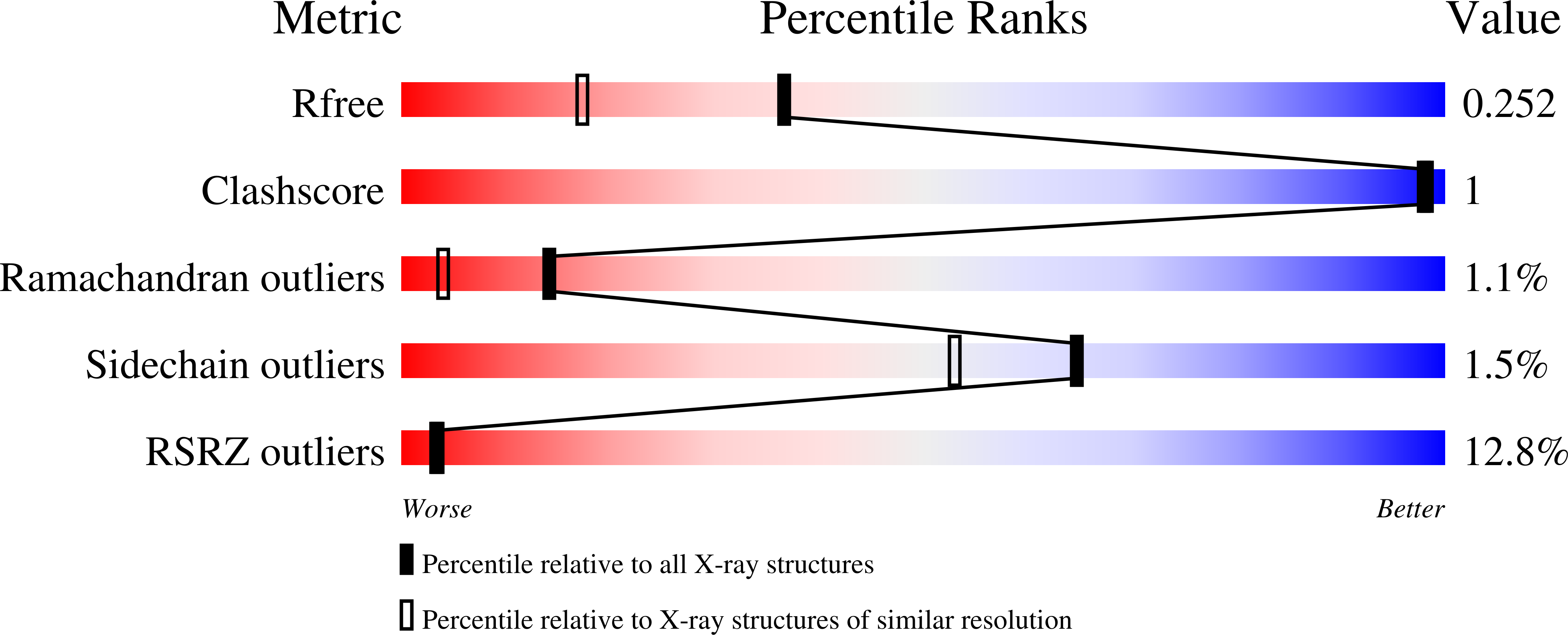
  • Method: X-RAY DIFFRACTION
  • Resolution: 1.85 Å
  • R-Value Free: 0.232 
  • R-Value Work: 0.217 
  • R-Value Observed: 0.217 

Starting Model: experimental
View more details

wwPDB Validation   3D Report Full Report


Ligand Structure Quality Assessment 


This is version 1.2 of the entry. See complete history


Literature

Using yeast surface display to engineer a soluble and crystallizable construct of hematopoietic progenitor kinase 1 (HPK1).

Lau, W.L.Pearce, B.Malakian, H.Rodrigo, I.Xie, D.Gao, M.Marsilio, F.Chang, C.Ruzanov, M.Muckelbauer, J.K.Newitt, J.A.Lipovsek, D.Sheriff, S.

(2021) Acta Crystallogr F Struct Biol Commun 77: 22-28

  • DOI: https://doi.org/10.1107/S2053230X20016015
  • Primary Citation of Related Structures:  
    7KAC

  • PubMed Abstract: 

    Hematopoietic progenitor kinase 1 (HPK1) is an intracellular kinase that plays an important role in modulating tumor immune response and thus is an attractive target for drug discovery. Crystallization of the wild-type HPK1 kinase domain has been hampered by poor expression in recombinant systems and poor solubility. In this study, yeast surface display was applied to a library of HPK1 kinase-domain variants in order to select variants with an improved expression level and solubility. The HPK1 variant with the most improved properties contained two mutations, crystallized readily in complex with several small-molecule inhibitors and provided valuable insight to guide structure-based drug design. This work exemplifies the benefit of yeast surface display towards engineering crystallizable proteins and thus enabling structure-based drug discovery.


  • Organizational Affiliation

    Biologics Discovery, Bristol-Myers Squibb Research and Development, 100 Binney Street, Cambridge, MA 02142, USA.


Macromolecules
Find similar proteins by:  (by identity cutoff)  |  3D Structure
Entity ID: 1
MoleculeChains Sequence LengthOrganismDetailsImage
Isoform 2 of Mitogen-activated protein kinase kinase kinase kinase 1
A, B
340Homo sapiensMutation(s): 2 
Gene Names: MAP4K1HPK1
EC: 2.7.11.1
UniProt & NIH Common Fund Data Resources
Find proteins for Q92918 (Homo sapiens)
Explore Q92918 
Go to UniProtKB:  Q92918
PHAROS:  Q92918
GTEx:  ENSG00000104814 
Entity Groups  
Sequence Clusters30% Identity50% Identity70% Identity90% Identity95% Identity100% Identity
UniProt GroupQ92918
Sequence Annotations
Expand
  • Reference Sequence
Small Molecules
Ligands 1 Unique
IDChains Name / Formula / InChI Key2D Diagram3D Interactions
W9D (Subject of Investigation/LOI)
Query on W9D

Download Ideal Coordinates CCD File 
C [auth A],
D [auth B]
5-{[4-{[(1S)-2-hydroxy-1-phenylethyl]amino}-5-(1,3,4-oxadiazol-2-yl)pyrimidin-2-yl]amino}-3,3-dimethyl-2-benzofuran-1(3H)-one
C24 H22 N6 O4
RVSSNRBUPQUIEG-LJQANCHMSA-N
Binding Affinity Annotations 
IDSourceBinding Affinity
W9D BindingDB:  7KAC IC50: min: 2.6, max: 290 (nM) from 2 assay(s)
Experimental Data & Validation

Experimental Data

  • Method: X-RAY DIFFRACTION
  • Resolution: 1.85 Å
  • R-Value Free: 0.232 
  • R-Value Work: 0.217 
  • R-Value Observed: 0.217 
  • Space Group: P 1
Unit Cell:
Length ( Å )Angle ( ˚ )
a = 55.83α = 78.81
b = 58.47β = 79.82
c = 60.48γ = 66.08
Software Package:
Software NamePurpose
XDSdata reduction
Aimlessdata scaling
AMoREphasing
BUSTERrefinement
PDB_EXTRACTdata extraction

Structure Validation

View Full Validation Report


Ligand Structure Quality Assessment 


Entry History 

Deposition Data

  • Released Date: 2021-01-13 
  • Deposition Author(s): Sheriff, S.

Revision History  (Full details and data files)

  • Version 1.0: 2021-01-13
    Type: Initial release
  • Version 1.1: 2021-01-27
    Changes: Database references
  • Version 1.2: 2023-10-18
    Changes: Data collection, Database references, Refinement description