6WP0

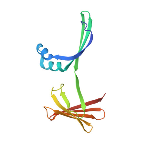
The Crystal Structure of Domain-Swapped Trimer Q108K:T51D variant of HCRBPII


Experimental Data Snapshot

  • Method: X-RAY DIFFRACTION
  • Resolution: 2.78 Å
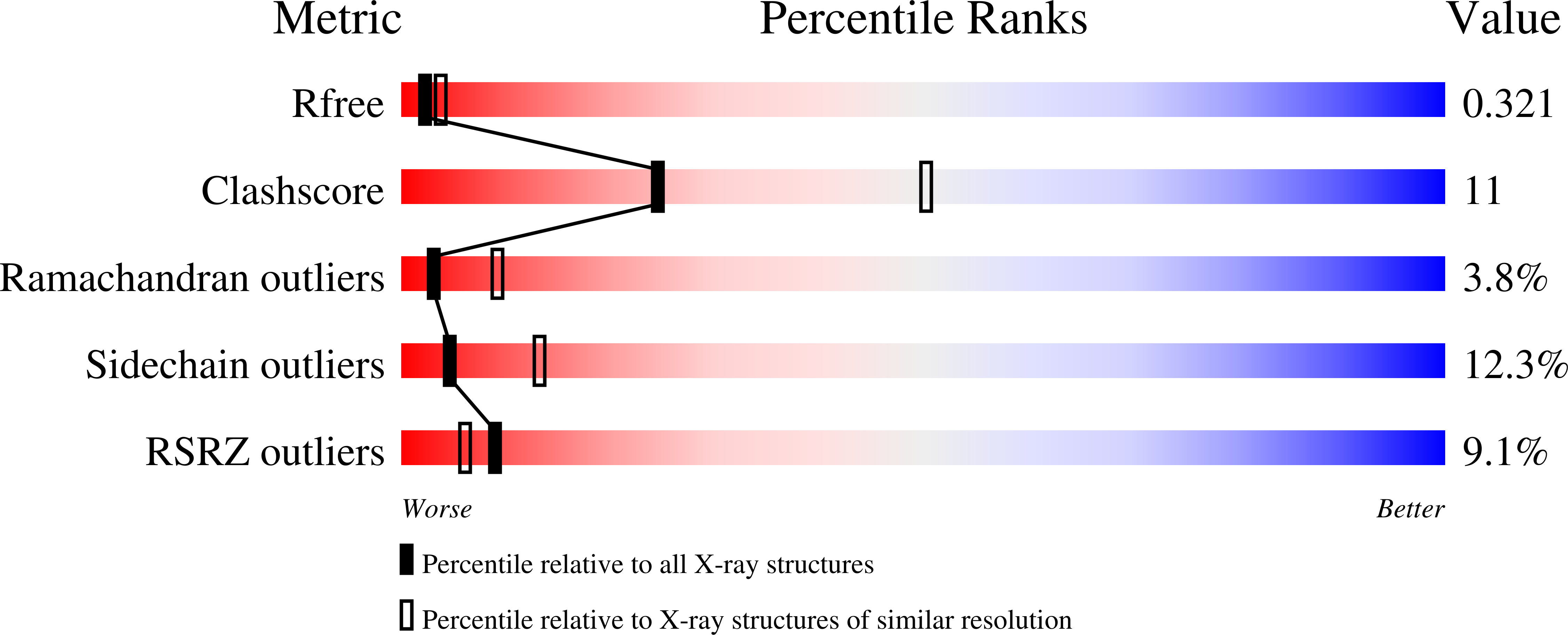
  • R-Value Free: 0.321 
  • R-Value Work: 0.276 
  • R-Value Observed: 0.278 

Starting Model: experimental
View more details

wwPDB Validation   3D Report Full Report


This is version 1.2 of the entry. See complete history


Literature

Human Cellular Retinol Binding Protein II Forms a Domain-Swapped Trimer Representing a Novel Fold and a New Template for Protein Engineering.

Ghanbarpour, A.Santos, E.M.Pinger, C.Assar, Z.Hossaini Nasr, S.Vasileiou, C.Spence, D.Borhan, B.Geiger, J.H.

(2020) Chembiochem 21: 3192-3196

  • DOI: https://doi.org/10.1002/cbic.202000405
  • Primary Citation of Related Structures:  
    6VIS, 6VIT, 6WNF, 6WNJ, 6WP0, 6WP1, 6WP2

  • PubMed Abstract: 

    Domain-swapping is a mechanism for evolving new protein structure from extant scaffolds, and has been an efficient protein-engineering strategy for tailoring functional diversity. However, domain swapping can only be exploited if it can be controlled, especially in cases where various folds can coexist. Herein, we describe the structure of a domain-swapped trimer of the iLBP family member hCRBPII, and suggest a mechanism for domain-swapped trimerization. It is further shown that domain-swapped trimerization can be favored by strategic installation of a disulfide bond, thus demonstrating a strategy for fold control. We further show the domain-swapped trimer to be a useful protein design template by installing a high-affinity metal binding site through the introduction of a single mutation, taking advantage of its threefold symmetry. Together, these studies show how nature can promote oligomerization, stabilize a specific oligomer, and generate new function with minimal changes to the protein sequence.


  • Organizational Affiliation

    Department of Chemistry, Michigan State University, 578 S. Shaw Lane, East Lansing, MI 48824, USA.


Macromolecules
Find similar proteins by:  (by identity cutoff)  |  3D Structure
Entity ID: 1
MoleculeChains Sequence LengthOrganismDetailsImage
Retinol-binding protein 2
A, B, C, D, E
A, B, C, D, E, F, G, H, I, J, K, L
133Homo sapiensMutation(s): 2 
Gene Names: RBP2CRBP2
UniProt & NIH Common Fund Data Resources
Find proteins for P50120 (Homo sapiens)
Explore P50120 
Go to UniProtKB:  P50120
PHAROS:  P50120
GTEx:  ENSG00000114113 
Entity Groups  
Sequence Clusters30% Identity50% Identity70% Identity90% Identity95% Identity100% Identity
UniProt GroupP50120
Sequence Annotations
Expand
  • Reference Sequence
Small Molecules
Ligands 2 Unique
IDChains Name / Formula / InChI Key2D Diagram3D Interactions
GOL
Query on GOL

Download Ideal Coordinates CCD File 
BA [auth J]
CA [auth J]
DA [auth J]
EA [auth J]
FA [auth K]
BA [auth J],
CA [auth J],
DA [auth J],
EA [auth J],
FA [auth K],
GA [auth K],
HA [auth L],
IA [auth L],
M [auth A],
N [auth A],
O [auth A],
P [auth A],
Q [auth B],
R [auth B],
S [auth B],
T [auth B],
V [auth D],
W [auth E],
X [auth E],
Y [auth G],
Z [auth H]
GLYCEROL
C3 H8 O3
PEDCQBHIVMGVHV-UHFFFAOYSA-N
ACT
Query on ACT

Download Ideal Coordinates CCD File 
AA [auth J],
U [auth D]
ACETATE ION
C2 H3 O2
QTBSBXVTEAMEQO-UHFFFAOYSA-M
Experimental Data & Validation

Experimental Data

  • Method: X-RAY DIFFRACTION
  • Resolution: 2.78 Å
  • R-Value Free: 0.321 
  • R-Value Work: 0.276 
  • R-Value Observed: 0.278 
  • Space Group: P 1 21 1
Unit Cell:
Length ( Å )Angle ( ˚ )
a = 84.565α = 90
b = 111.272β = 112.89
c = 101.826γ = 90
Software Package:
Software NamePurpose
PHENIXrefinement
PHENIXrefinement
HKL-2000data reduction
HKL-2000data scaling
PHASERphasing

Structure Validation

View Full Validation Report


Entry History & Funding Information

Deposition Data


Funding OrganizationLocationGrant Number
National Institutes of Health/National Institute of General Medical Sciences (NIH/NIGMS)United StatesGM101353

Revision History  (Full details and data files)

  • Version 1.0: 2020-08-19
    Type: Initial release
  • Version 1.1: 2021-05-05
    Changes: Database references
  • Version 1.2: 2023-10-18
    Changes: Data collection, Database references, Refinement description