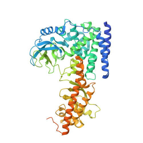
6V62

SETD3 double mutant (N255F/W273A) in Complex with an Actin Peptide with His73 Replaced with Lysine


Experimental Data Snapshot

  • Method: X-RAY DIFFRACTION
  • Resolution: 2.36 Å
  • R-Value Free: 0.252 
  • R-Value Work: 0.208 
  • R-Value Observed: 0.210 

Starting Model: experimental
View more details

wwPDB Validation   3D Report Full Report

Currently 6V62 does not have a validation slider image.


This is version 1.3 of the entry. See complete history


Literature

An engineered variant of SETD3 methyltransferase alters target specificity from histidine to lysine methylation.

Dai, S.Horton, J.R.Wilkinson, A.W.Gozani, O.Zhang, X.Cheng, X.

(2020) J Biol Chem 295: 2582-2589

  • DOI: https://doi.org/10.1074/jbc.RA119.012319
  • Primary Citation of Related Structures:  
    6V62, 6V63

  • PubMed Abstract: 

    Most characterized SET domain (SETD) proteins are protein lysine methyltransferases, but SETD3 was recently demonstrated to be a protein ( i.e. actin) histidine-N 3 methyltransferase. Human SETD3 shares a high structural homology with two known protein lysine methyltransferases-human SETD6 and the plant LSMT-but differs in the residues constituting the active site. In the SETD3 active site, Asn 255 engages in a unique hydrogen-bonding interaction with the target histidine of actin that likely contributes to its >1300-fold greater catalytic efficiency ( k cat / K m ) on histidine than on lysine. Here, we engineered active-site variants to switch the SETD3 target specificity from histidine to lysine. Substitution of Asn 255 with phenylalanine (N255F), together with substitution of Trp 273 with alanine (W273A), generated an active site mimicking that of known lysine methyltransferases. The doubly substituted SETD3 variant exhibited a 13-fold preference for lysine over histidine. We show, by means of X-ray crystallography, that the two target nitrogen atoms-the N 3 atom of histidine and the terminal ϵ-amino nitrogen of lysine-occupy the same position and point toward and are within a short distance of the incoming methyl group of SAM for a direct methyl transfer during catalysis. In contrast, SETD3 and its Asn 255 substituted derivatives did not methylate glutamine (another potentially methylated amino acid). However, the glutamine-containing peptide competed with the substrate peptide, and glutamine bound in the active site, but too far away from SAM to be methylated. Our results provide insight into the structural parameters defining the target amino acid specificity of SET enzymes.


  • Organizational Affiliation

    Department of Epigenetics and Molecular Carcinogenesis, University of Texas M.D. Anderson Cancer Center, Houston, Texas 77030.


Macromolecules

Find similar proteins by:  Sequence   |   3D Structure  

Entity ID: 1
MoleculeChains Sequence LengthOrganismDetailsImage
Actin, cytoplasmic 1A [auth Y]23Homo sapiensMutation(s): 1 
EC: 3.6.4
UniProt & NIH Common Fund Data Resources
Find proteins for P63261 (Homo sapiens)
Explore P63261 
Go to UniProtKB:  P63261
PHAROS:  P63261
GTEx:  ENSG00000184009 
Entity Groups  
Sequence Clusters30% Identity50% Identity70% Identity90% Identity95% Identity100% Identity
UniProt GroupP63261
Sequence Annotations
Expand
  • Reference Sequence
Find similar proteins by:  (by identity cutoff)  |  3D Structure
Entity ID: 2
MoleculeChains Sequence LengthOrganismDetailsImage
Actin-histidine N-methyltransferaseB [auth A]596Homo sapiensMutation(s): 2 
Gene Names: SETD3C14orf154
EC: 2.1.1.85
UniProt & NIH Common Fund Data Resources
Find proteins for Q86TU7 (Homo sapiens)
Explore Q86TU7 
Go to UniProtKB:  Q86TU7
PHAROS:  Q86TU7
GTEx:  ENSG00000183576 
Entity Groups  
Sequence Clusters30% Identity50% Identity70% Identity90% Identity95% Identity100% Identity
UniProt GroupQ86TU7
Sequence Annotations
Expand
  • Reference Sequence
Experimental Data & Validation

Experimental Data

  • Method: X-RAY DIFFRACTION
  • Resolution: 2.36 Å
  • R-Value Free: 0.252 
  • R-Value Work: 0.208 
  • R-Value Observed: 0.210 
  • Space Group: C 2 2 21
Unit Cell:
Length ( Å )Angle ( ˚ )
a = 60.654α = 90
b = 116.752β = 90
c = 174.377γ = 90
Software Package:
Software NamePurpose
PHENIXrefinement
HKL-2000data reduction
HKL-2000data scaling
PHENIXphasing

Structure Validation

View Full Validation Report

Currently 6V62 does not have a validation slider image.


Entry History & Funding Information

Deposition Data


Funding OrganizationLocationGrant Number
National Institutes of Health/National Institute of General Medical Sciences (NIH/NIGMS)United StatesGM049245-23

Revision History  (Full details and data files)

  • Version 1.0: 2020-01-15
    Type: Initial release
  • Version 1.1: 2020-01-22
    Changes: Database references
  • Version 1.2: 2020-03-11
    Changes: Database references
  • Version 1.3: 2023-10-11
    Changes: Data collection, Database references, Refinement description