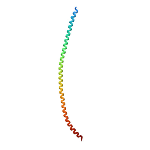
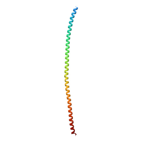
6UUI

Crystal structure of the heterocomplex between coil 2B domains of wild-type keratin 1 (KRT1) and keratin 10 (KRT10) containing mutation Cys401Ala


Experimental Data Snapshot

  • Method: X-RAY DIFFRACTION
  • Resolution: 2.07 Å
  • R-Value Free: 0.277 
  • R-Value Work: 0.250 
  • R-Value Observed: 0.252 

Starting Model: experimental
View more details

wwPDB Validation   3D Report Full Report

Currently 6UUI does not have a validation slider image.


This is version 1.4 of the entry. See complete history


Literature

Crystal Structure of Keratin 1/10(C401A) 2B Heterodimer Demonstrates a Proclivity for the C-Terminus of Helix 2B to Form Higher Order Molecular Contacts.

Lomakin, I.B.Hinbest, A.J.Ho, M.Eldirany, S.A.Bunick, C.G.

(2020) Yale J Biol Med 93: 3-17

  • Primary Citation of Related Structures:  
    6UUI

  • PubMed Abstract: 

    We previously determined the crystal structure of the wild-type keratin 1/10 helix 2B heterodimer at 3.3 Å resolution. We proposed that the resolution of the diffraction data was limited due to the crystal packing effect from keratin 10 (K10) residue Cys401. Cys401 K10 formed a disulfide-linkage with Cys401 from another K1/10 heterodimer, creating an "X-shaped" structure and a loose crystal packing arrangement. We hypothesized that mutation of Cys401 K10 to alanine would eliminate the disulfide-linkage and improve crystal packing thereby increasing resolution of diffraction and enabling a more accurate side chain electron density map. Indeed, when a K10 Cys401Ala 2B mutant was paired with its native keratin 1 (K1) 2B heterodimer partner its x-ray crystal structure was determined at 2.07 Å resolution; the structure does not contain a disulfide linkage. Superposition of the K1/K10(Cys401Ala) 2B structure onto the wild-type K1/10 2B heterodimer structure had a root-mean-square-deviation of 1.88 Å; the variability in the atomic positions reflects the dynamic motion expected in this filamentous coiled-coil complex. The electrostatic, hydrophobic, and contour features of the molecular surface are similar to the lower resolution wild-type structure. We postulated that elimination of the disulfide linkage in the K1/K10(Cys401Ala) 2B structure could allow for the 2B heterodimers to bind/pack in the A 22 tetramer configuration associated with mature keratin intermediate filament assembly. Analysis of the crystal packing revealed a half-staggered anti-parallel tetrameric complex of 2B heterodimers; however, their register is not consistent with models of the A 22 mode of tetrameric alignment or prior biochemical cross-linking studies.


  • Organizational Affiliation

    Department of Molecular Biophysics and Biochemistry, Yale University, New Haven, CT.


Macromolecules
Find similar proteins by:  (by identity cutoff)  |  3D Structure
Entity ID: 1
MoleculeChains Sequence LengthOrganismDetailsImage
Keratin, type II cytoskeletal 1A [auth C]120Homo sapiensMutation(s): 0 
Gene Names: KRT1KRTA
UniProt & NIH Common Fund Data Resources
Find proteins for P04264 (Homo sapiens)
Explore P04264 
Go to UniProtKB:  P04264
PHAROS:  P04264
GTEx:  ENSG00000167768 
Entity Groups  
Sequence Clusters30% Identity50% Identity70% Identity90% Identity95% Identity100% Identity
UniProt GroupP04264
Sequence Annotations
Expand
  • Reference Sequence
Find similar proteins by:  (by identity cutoff)  |  3D Structure
Entity ID: 2
MoleculeChains Sequence LengthOrganismDetailsImage
Keratin, type I cytoskeletal 10B [auth X]122Homo sapiensMutation(s): 1 
Gene Names: KRT10KPP
UniProt & NIH Common Fund Data Resources
Find proteins for P13645 (Homo sapiens)
Explore P13645 
Go to UniProtKB:  P13645
PHAROS:  P13645
GTEx:  ENSG00000186395 
Entity Groups  
Sequence Clusters30% Identity50% Identity70% Identity90% Identity95% Identity100% Identity
UniProt GroupP13645
Sequence Annotations
Expand
  • Reference Sequence
Small Molecules
Ligands 2 Unique
IDChains Name / Formula / InChI Key2D Diagram3D Interactions
BOG
Query on BOG

Download Ideal Coordinates CCD File 
C
octyl beta-D-glucopyranoside
C14 H28 O6
HEGSGKPQLMEBJL-RKQHYHRCSA-N
GOL
Query on GOL

Download Ideal Coordinates CCD File 
D [auth X]GLYCEROL
C3 H8 O3
PEDCQBHIVMGVHV-UHFFFAOYSA-N
Experimental Data & Validation

Experimental Data

  • Method: X-RAY DIFFRACTION
  • Resolution: 2.07 Å
  • R-Value Free: 0.277 
  • R-Value Work: 0.250 
  • R-Value Observed: 0.252 
  • Space Group: P 61 2 2
Unit Cell:
Length ( Å )Angle ( ˚ )
a = 100.124α = 90
b = 100.124β = 90
c = 224.788γ = 120
Software Package:
Software NamePurpose
PHENIXrefinement
HKL-2000data reduction
HKL-2000data scaling
MOLREPphasing
PDB_EXTRACTdata extraction

Structure Validation

View Full Validation Report

Currently 6UUI does not have a validation slider image.


Entry History & Funding Information

Deposition Data


Funding OrganizationLocationGrant Number
National Institutes of Health/National Institute of Arthritis and Musculoskeletal and Skin Diseases (NIH/NIAMS)United StatesK08AR070290
Other privateUnited StatesFoundation for Ichthyosis and Related Skin Types

Revision History  (Full details and data files)

  • Version 1.0: 2019-11-13
    Type: Initial release
  • Version 1.1: 2019-12-11
    Changes: Author supporting evidence, Database references
  • Version 1.2: 2020-04-08
    Changes: Database references
  • Version 1.3: 2020-07-29
    Type: Remediation
    Reason: Carbohydrate remediation
    Changes: Data collection, Derived calculations, Structure summary
  • Version 1.4: 2023-10-11
    Changes: Data collection, Database references, Refinement description, Structure summary