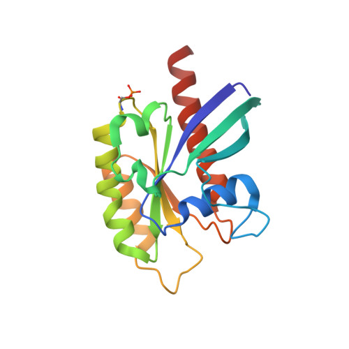
6STF

Human Rab8a phosphorylated at Ser111 in complex with GDP


Experimental Data Snapshot

  • Method: X-RAY DIFFRACTION
  • Resolution: 2.40 Å
  • R-Value Free: 0.262 
  • R-Value Work: 0.233 
  • R-Value Observed: 0.234 

Starting Model: experimental
View more details

wwPDB Validation   3D Report Full Report


Ligand Structure Quality Assessment 


This is version 1.3 of the entry. See complete history


Literature

PINK1-dependent phosphorylation of Serine111 within the SF3 motif of Rab GTPases impairs effector interactions and LRRK2-mediated phosphorylation at Threonine72.

Vieweg, S.Mulholland, K.Brauning, B.Kachariya, N.Lai, Y.C.Toth, R.Singh, P.K.Volpi, I.Sattler, M.Groll, M.Itzen, A.Muqit, M.M.K.

(2020) Biochem J 477: 1651-1668

  • DOI: https://doi.org/10.1042/BCJ20190664
  • Primary Citation of Related Structures:  
    6STF, 6STG

  • PubMed Abstract: 

    Loss of function mutations in the PTEN-induced kinase 1 (PINK1) kinase are causal for autosomal recessive Parkinson's disease (PD) whilst gain of function mutations in the LRRK2 kinase cause autosomal dominant PD. PINK1 indirectly regulates the phosphorylation of a subset of Rab GTPases at a conserved Serine111 (Ser111) residue within the SF3 motif. Using genetic code expansion technologies, we have produced stoichiometric Ser111-phosphorylated Rab8A revealing impaired interactions with its cognate guanine nucleotide exchange factor and GTPase activating protein. In a screen for Rab8A kinases we identify TAK1 and MST3 kinases that can efficiently phosphorylate the Switch II residue Threonine72 (Thr72) in a similar manner as LRRK2 in vitro. Strikingly, we demonstrate that Ser111 phosphorylation negatively regulates the ability of LRRK2 but not MST3 or TAK1 to phosphorylate Thr72 of recombinant nucleotide-bound Rab8A in vitro and demonstrate an interplay of PINK1- and LRRK2-mediated phosphorylation of Rab8A in transfected HEK293 cells. Finally, we present the crystal structure of Ser111-phosphorylated Rab8A and nuclear magnetic resonance structure of Ser111-phosphorylated Rab1B. The structures reveal that the phosphorylated SF3 motif does not induce any major changes, but may interfere with effector-Switch II interactions through intramolecular H-bond formation and/or charge effects with Arg79. Overall, we demonstrate antagonistic regulation between PINK1-dependent Ser111 phosphorylation and LRRK2-mediated Thr72 phosphorylation of Rab8A indicating a potential cross-talk between PINK1-regulated mitochondrial homeostasis and LRRK2 signalling that requires further investigation in vivo.


  • Organizational Affiliation

    Department of Chemistry, Centre for Integrated Protein Science Munich (CIPSM), Technical University of Munich, Garching 85748, Germany.


Macromolecules
Find similar proteins by:  (by identity cutoff)  |  3D Structure
Entity ID: 1
MoleculeChains Sequence LengthOrganismDetailsImage
Ras-related protein Rab-8A
A, B, C, D, E
178Homo sapiensMutation(s): 0 
Gene Names: RAB8AMELRAB8
EC: 3.6.5.2
UniProt & NIH Common Fund Data Resources
Find proteins for P61006 (Homo sapiens)
Explore P61006 
Go to UniProtKB:  P61006
PHAROS:  P61006
GTEx:  ENSG00000167461 
Entity Groups  
Sequence Clusters30% Identity50% Identity70% Identity90% Identity95% Identity100% Identity
UniProt GroupP61006
Sequence Annotations
Expand
  • Reference Sequence
Small Molecules
Ligands 2 Unique
IDChains Name / Formula / InChI Key2D Diagram3D Interactions
GDP (Subject of Investigation/LOI)
Query on GDP

Download Ideal Coordinates CCD File 
F [auth A],
H [auth B],
J [auth C],
L [auth D],
N [auth E]
GUANOSINE-5'-DIPHOSPHATE
C10 H15 N5 O11 P2
QGWNDRXFNXRZMB-UUOKFMHZSA-N
MG (Subject of Investigation/LOI)
Query on MG

Download Ideal Coordinates CCD File 
G [auth A],
I [auth B],
K [auth C],
M [auth D],
O [auth E]
MAGNESIUM ION
Mg
JLVVSXFLKOJNIY-UHFFFAOYSA-N
Modified Residues  1 Unique
IDChains TypeFormula2D DiagramParent
SEP
Query on SEP
A, B, C, D, E
L-PEPTIDE LINKINGC3 H8 N O6 PSER
Experimental Data & Validation

Experimental Data

  • Method: X-RAY DIFFRACTION
  • Resolution: 2.40 Å
  • R-Value Free: 0.262 
  • R-Value Work: 0.233 
  • R-Value Observed: 0.234 
  • Space Group: C 1 2 1
Unit Cell:
Length ( Å )Angle ( ˚ )
a = 117.55α = 90
b = 75.18β = 100.48
c = 107.7γ = 90
Software Package:
Software NamePurpose
REFMACrefinement
PDB_EXTRACTdata extraction
XDSdata reduction
XSCALEdata scaling
PHASERphasing

Structure Validation

View Full Validation Report


Ligand Structure Quality Assessment 


Entry History & Funding Information

Deposition Data


Funding OrganizationLocationGrant Number
German Research FoundationGermanySFB1035

Revision History  (Full details and data files)

  • Version 1.0: 2020-04-22
    Type: Initial release
  • Version 1.1: 2020-05-20
    Changes: Database references
  • Version 1.2: 2024-01-24
    Changes: Data collection, Database references, Derived calculations, Refinement description
  • Version 1.3: 2024-10-16
    Changes: Structure summary