6SGE

Crystal structure of Human RHOB-GTP in complex with nanobody B6


Experimental Data Snapshot

  • Method: X-RAY DIFFRACTION
  • Resolution: 1.50 Å
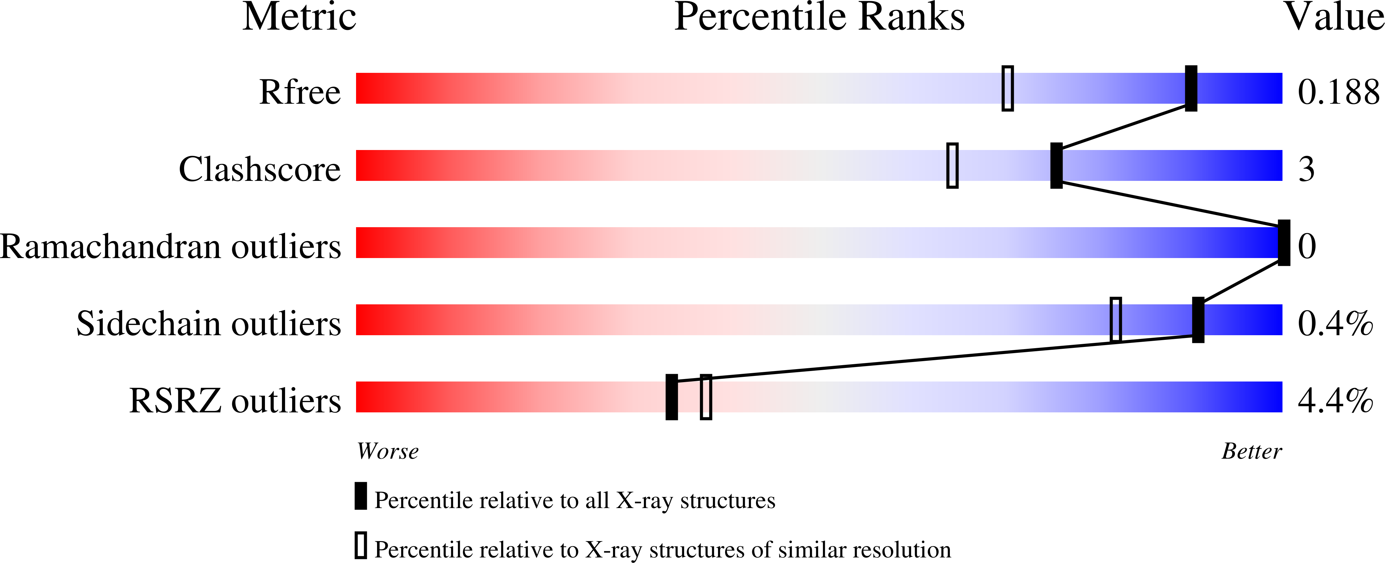
  • R-Value Free: 0.188 
  • R-Value Work: 0.161 
  • R-Value Observed: 0.162 

Starting Model: experimental
View more details

wwPDB Validation   3D Report Full Report


Ligand Structure Quality Assessment 


This is version 1.2 of the entry. See complete history


Literature

A Targeted Protein Degradation Cell-Based Screening for Nanobodies Selective toward the Cellular RHOB GTP-Bound Conformation.

Bery, N.Keller, L.Soulie, M.Gence, R.Iscache, A.L.Cherier, J.Cabantous, S.Sordet, O.Lajoie-Mazenc, I.Pedelacq, J.D.Favre, G.Olichon, A.

(2019) Cell Chem Biol 26: 1544

  • DOI: https://doi.org/10.1016/j.chembiol.2019.08.009
  • Primary Citation of Related Structures:  
    6HXU, 6SGE

  • PubMed Abstract: 

    The selective downregulation of activated intracellular proteins is a key challenge in cell biology. RHO small GTPases switch between a guanosine diphosphate (GDP)-bound and a guanosine triphosphate (GTP)-bound state that drives downstream signaling. At present, no tool is available to study endogenous RHO-GTPinduced conformational changes in live cells. Here, we established a cell-based screen to selectively degrade RHOB-GTP using F-box-intracellular single-domain antibody fusion. We identified one intracellular antibody (intrabody) that shows selective targeting of endogenous RHOB-GTP mediated by interactions between the CDR3 loop of the domain antibody and the GTP-binding pocket of RHOB. Our results suggest that, while RHOB is highly regulated at the expression level, only the GTP-bound pool, but not its global expression, mediates RHOB functions in genomic instability and in cell invasion. The F-box/intrabody-targeted protein degradation represents a unique approach to knock down the active form of small GTPases or other proteins with multiple cellular activities.


  • Organizational Affiliation

    Centre de Recherche en Cancérologie de Toulouse (CRCT), INSERM, Université de Toulouse, CNRS, UPS, Toulouse, France.


Macromolecules
Find similar proteins by:  (by identity cutoff)  |  3D Structure
Entity ID: 1
MoleculeChains Sequence LengthOrganismDetailsImage
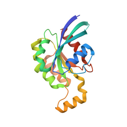
Rho-related GTP-binding protein RhoB
A, C
183Homo sapiensMutation(s): 1 
Gene Names: RHOBARH6ARHB
UniProt & NIH Common Fund Data Resources
Find proteins for P62745 (Homo sapiens)
Explore P62745 
Go to UniProtKB:  P62745
PHAROS:  P62745
GTEx:  ENSG00000143878 
Entity Groups  
Sequence Clusters30% Identity50% Identity70% Identity90% Identity95% Identity100% Identity
UniProt GroupP62745
Sequence Annotations
Expand
  • Reference Sequence
Find similar proteins by:  (by identity cutoff)  |  3D Structure
Entity ID: 2
MoleculeChains Sequence LengthOrganismDetailsImage
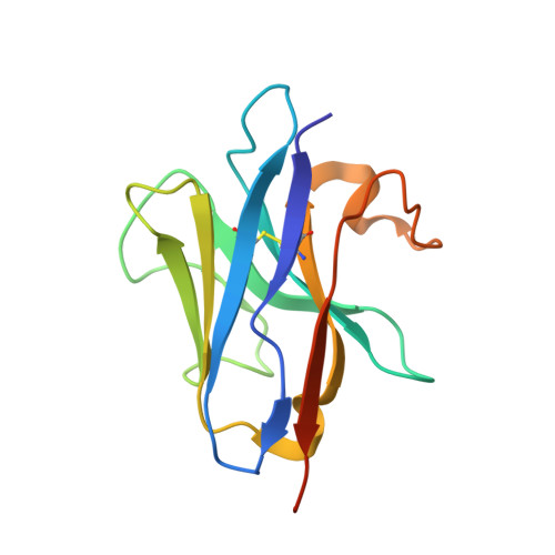
Nanobody B6
B, D
134Homo sapiensMutation(s): 0 
UniProt
Find proteins for A0A4E0W6L3 (Homo sapiens)
Explore A0A4E0W6L3 
Go to UniProtKB:  A0A4E0W6L3
Entity Groups  
Sequence Clusters30% Identity50% Identity70% Identity90% Identity95% Identity100% Identity
UniProt GroupA0A4E0W6L3
Sequence Annotations
Expand
  • Reference Sequence
Experimental Data & Validation

Experimental Data

  • Method: X-RAY DIFFRACTION
  • Resolution: 1.50 Å
  • R-Value Free: 0.188 
  • R-Value Work: 0.161 
  • R-Value Observed: 0.162 
  • Space Group: P 1 21 1
Unit Cell:
Length ( Å )Angle ( ˚ )
a = 66.84α = 90
b = 70.227β = 107.68
c = 71.212γ = 90
Software Package:
Software NamePurpose
PHENIXrefinement
XDSdata reduction
XDSdata scaling
PHENIXphasing
PDB_EXTRACTdata extraction

Structure Validation

View Full Validation Report


Ligand Structure Quality Assessment 


Entry History 

Deposition Data

Revision History  (Full details and data files)

  • Version 1.0: 2019-09-25
    Type: Initial release
  • Version 1.1: 2019-12-04
    Changes: Database references
  • Version 1.2: 2024-01-24
    Changes: Data collection, Database references, Derived calculations, Refinement description