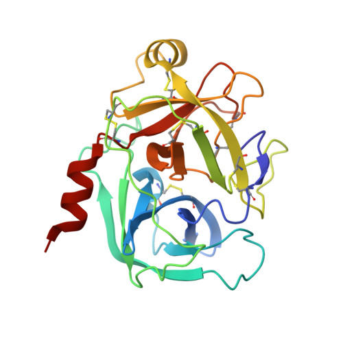
6QHB

Crystal Structure of Human Kallikrein 6 in complex with GSK578724A


Experimental Data Snapshot

  • Method: X-RAY DIFFRACTION
  • Resolution: 1.84 Å
  • R-Value Free: 0.206 
  • R-Value Work: 0.183 
  • R-Value Observed: 0.185 

Starting Model: experimental
View more details

wwPDB Validation   3D Report Full Report


Ligand Structure Quality Assessment 


This is version 1.2 of the entry. See complete history


Literature

Kallikrein 5 inhibitors identified through structure based drug design in search for a treatment for Netherton Syndrome.

White, G.V.Edgar, E.V.Holmes, D.S.Lewell, X.Q.Liddle, J.Polyakova, O.Smith, K.J.Thorpe, J.H.Walker, A.L.Wang, Y.Young, R.J.Hovnanian, A.

(2019) Bioorg Med Chem Lett 29: 821-825

  • DOI: https://doi.org/10.1016/j.bmcl.2019.01.020
  • Primary Citation of Related Structures:  
    6QH9, 6QHA, 6QHB, 6QHC

  • PubMed Abstract: 

    Netherton syndrome (NS) is a rare and debilitating severe autosomal recessive genetic skin disease with high mortality rates particularly in neonates. NS is caused by loss-of-function SPINK5 mutations leading to unregulated kallikrein 5 (KLK5) and kallikrein 7 (KLK7) activity. Furthermore, KLK5 inhibition has been proposed as a potential therapeutic treatment for NS. Identification of potent and selective KLK5 inhibitors would enable further exploration of the disease biology and could ultimately lead to a treatment for NS. This publication describes how fragmentation of known trypsin-like serine protease (TLSP) inhibitors resulted in the identification of a series of phenolic amidine-based KLK5 inhibitors 1. X-ray crystallography was used to find alternatives to the phenol interaction leading to identification of carbonyl analogues such as lactam 13 and benzimidazole 15. These reversible inhibitors, with selectivity over KLK1 (10-100 fold), provided novel starting points for the guided growth towards suitable tool molecules for the exploration of KLK5 biology.


  • Organizational Affiliation

    GlaxoSmithKline R&D, Medicines Research Centre, Gunnels Wood Road, Stevenage, Hertfordshire SG1 2NY, UK. Electronic address: gemma.v.white@gsk.com.


Macromolecules
Find similar proteins by:  (by identity cutoff)  |  3D Structure
Entity ID: 1
MoleculeChains Sequence LengthOrganismDetailsImage
Kallikrein-6
A, B
223Homo sapiensMutation(s): 3 
Gene Names: KLK6PRSS18PRSS9
EC: 3.4.21
UniProt & NIH Common Fund Data Resources
Find proteins for Q92876 (Homo sapiens)
Explore Q92876 
Go to UniProtKB:  Q92876
PHAROS:  Q92876
GTEx:  ENSG00000167755 
Entity Groups  
Sequence Clusters30% Identity50% Identity70% Identity90% Identity95% Identity100% Identity
UniProt GroupQ92876
Sequence Annotations
Expand
  • Reference Sequence
Small Molecules
Ligands 2 Unique
IDChains Name / Formula / InChI Key2D Diagram3D Interactions
J2W
Query on J2W

Download Ideal Coordinates CCD File 
C [auth A],
E [auth B]
~{N}-(4-carbamimidoylphenyl)-3-methoxy-2-oxidanyl-benzamide
C15 H15 N3 O3
FRAFAKWXNNKJCF-UHFFFAOYSA-N
GOL
Query on GOL

Download Ideal Coordinates CCD File 
D [auth B]GLYCEROL
C3 H8 O3
PEDCQBHIVMGVHV-UHFFFAOYSA-N
Experimental Data & Validation

Experimental Data

  • Method: X-RAY DIFFRACTION
  • Resolution: 1.84 Å
  • R-Value Free: 0.206 
  • R-Value Work: 0.183 
  • R-Value Observed: 0.185 
  • Space Group: P 1 21 1
Unit Cell:
Length ( Å )Angle ( ˚ )
a = 57.84α = 90
b = 45.95β = 93.54
c = 90.73γ = 90
Software Package:
Software NamePurpose
BUSTERrefinement
XDSdata reduction
Aimlessdata scaling
PHASERphasing

Structure Validation

View Full Validation Report


Ligand Structure Quality Assessment 


Entry History 

Deposition Data

  • Released Date: 2019-02-06 
  • Deposition Author(s): Thorpe, J.H.

Revision History  (Full details and data files)

  • Version 1.0: 2019-02-06
    Type: Initial release
  • Version 1.1: 2019-02-27
    Changes: Data collection, Database references
  • Version 1.2: 2024-01-24
    Changes: Data collection, Database references, Refinement description