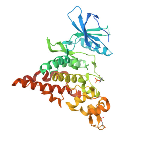
6P5S

HIPK2 kinase domain bound to CX-4945


Experimental Data Snapshot

  • Method: X-RAY DIFFRACTION
  • Resolution: 2.19 Å
  • R-Value Free: 0.238 
  • R-Value Work: 0.198 
  • R-Value Observed: 0.200 

wwPDB Validation   3D Report Full Report


Ligand Structure Quality Assessment 


This is version 1.2 of the entry. See complete history


Literature

The crystal structure of the protein kinase HIPK2 reveals a unique architecture of its CMGC-insert region.

Agnew, C.Liu, L.Liu, S.Xu, W.You, L.Yeung, W.Kannan, N.Jablons, D.Jura, N.

(2019) J Biol Chem 294: 13545-13559

  • DOI: https://doi.org/10.1074/jbc.RA119.009725
  • Primary Citation of Related Structures:  
    6P5S

  • PubMed Abstract: 

    The homeodomain-interacting protein kinase (HIPK) family is comprised of four nuclear protein kinases, HIPK1-4. HIPK proteins phosphorylate a diverse range of transcription factors involved in cell proliferation, differentiation, and apoptosis. HIPK2, thus far the best-characterized member of this largely understudied family of protein kinases, plays a role in the activation of p53 in response to DNA damage. Despite this tumor-suppressor function, HIPK2 is also found overexpressed in several cancers, and its hyperactivation causes chronic fibrosis. There are currently no structures of HIPK2 or of any other HIPK kinase. Here, we report the crystal structure of HIPK2's kinase domain bound to CX-4945, a casein kinase 2α (CK2α) inhibitor currently in clinical trials against several cancers. The structure, determined at 2.2 Å resolution, revealed that CX-4945 engages the HIPK2 active site in a hybrid binding mode between that seen in structures of CK2α and Pim1 kinases. The HIPK2 kinase domain crystallized in the active conformation, which was stabilized by phosphorylation of the activation loop. We noted that the overall kinase domain fold of HIPK2 closely resembles that of evolutionarily related dual-specificity tyrosine-regulated kinases (DYRKs). Most significant structural differences between HIPK2 and DYRKs included an absence of the regulatory N-terminal domain and a unique conformation of the CMGC-insert region and of a newly defined insert segment in the αC-β4 loop. This first crystal structure of HIPK2 paves the way for characterizing the understudied members of the HIPK family and for developing HIPK2-directed therapies for managing cancer and fibrosis.


  • Organizational Affiliation

    Cardiovascular Research Institute, University of California San Francisco, San Francisco, California 94158.


Macromolecules
Find similar proteins by:  (by identity cutoff)  |  3D Structure
Entity ID: 1
MoleculeChains Sequence LengthOrganismDetailsImage
Homeodomain-interacting protein kinase 2390Homo sapiensMutation(s): 0 
Gene Names: HIPK2
EC: 2.7.11.1
UniProt & NIH Common Fund Data Resources
Find proteins for Q9H2X6 (Homo sapiens)
Explore Q9H2X6 
Go to UniProtKB:  Q9H2X6
PHAROS:  Q9H2X6
GTEx:  ENSG00000064393 
Entity Groups  
Sequence Clusters30% Identity50% Identity70% Identity90% Identity95% Identity100% Identity
UniProt GroupQ9H2X6
Sequence Annotations
Expand
  • Reference Sequence
Small Molecules
Ligands 1 Unique
IDChains Name / Formula / InChI Key2D Diagram3D Interactions
3NG
Query on 3NG

Download Ideal Coordinates CCD File 
B [auth A]5-[(3-chlorophenyl)amino]benzo[c][2,6]naphthyridine-8-carboxylic acid
C19 H12 Cl N3 O2
MUOKSQABCJCOPU-UHFFFAOYSA-N
Modified Residues  2 Unique
IDChains TypeFormula2D DiagramParent
PTR
Query on PTR
A
L-PEPTIDE LINKINGC9 H12 N O6 PTYR
SEP
Query on SEP
A
L-PEPTIDE LINKINGC3 H8 N O6 PSER
Binding Affinity Annotations 
IDSourceBinding Affinity
3NG BindingDB:  6P5S IC50: 45 (nM) from 1 assay(s)
Experimental Data & Validation

Experimental Data

  • Method: X-RAY DIFFRACTION
  • Resolution: 2.19 Å
  • R-Value Free: 0.238 
  • R-Value Work: 0.198 
  • R-Value Observed: 0.200 
  • Space Group: P 62
Unit Cell:
Length ( Å )Angle ( ˚ )
a = 130.155α = 90
b = 130.155β = 90
c = 52.253γ = 120
Software Package:
Software NamePurpose
PHENIXrefinement
HKL-2000data reduction
SCALEPACKdata scaling
PHASERphasing

Structure Validation

View Full Validation Report


Ligand Structure Quality Assessment 


Entry History 

Deposition Data

Revision History  (Full details and data files)

  • Version 1.0: 2019-07-31
    Type: Initial release
  • Version 1.1: 2019-08-07
    Changes: Data collection, Database references
  • Version 1.2: 2019-09-25
    Changes: Data collection, Database references