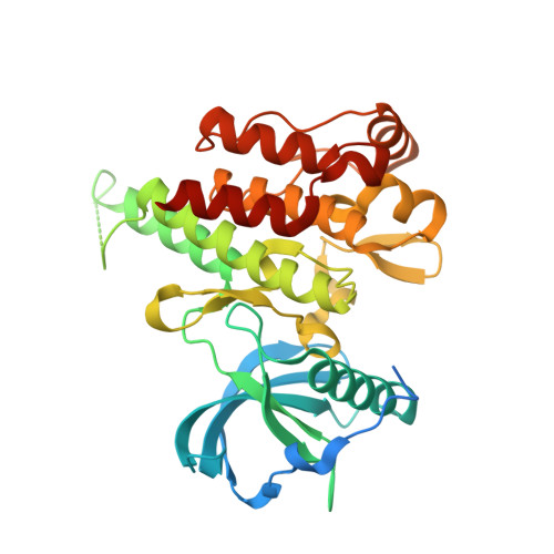
6N33

Crystal structure of fms kinase domain with a small molecular inhibitor, PLX5622


Experimental Data Snapshot

  • Method: X-RAY DIFFRACTION
  • Resolution: 2.25 Å
  • R-Value Free: 0.220 
  • R-Value Work: 0.181 
  • R-Value Observed: 0.183 

Starting Model: experimental
View more details

wwPDB Validation   3D Report Full Report


Ligand Structure Quality Assessment 


This is version 1.1 of the entry. See complete history


Literature

Sustained microglial depletion with CSF1R inhibitor impairs parenchymal plaque development in an Alzheimer's disease model.

Spangenberg, E.Severson, P.L.Hohsfield, L.A.Crapser, J.Zhang, J.Burton, E.A.Zhang, Y.Spevak, W.Lin, J.Phan, N.Y.Habets, G.Rymar, A.Tsang, G.Walters, J.Nespi, M.Singh, P.Broome, S.Ibrahim, P.Zhang, C.Bollag, G.West, B.L.Green, K.N.

(2019) Nat Commun 10: 3758-3758

  • DOI: https://doi.org/10.1038/s41467-019-11674-z
  • Primary Citation of Related Structures:  
    6N33

  • PubMed Abstract: 

    Many risk genes for the development of Alzheimer's disease (AD) are exclusively or highly expressed in myeloid cells. Microglia are dependent on colony-stimulating factor 1 receptor (CSF1R) signaling for their survival. We designed and synthesized a highly selective brain-penetrant CSF1R inhibitor (PLX5622) allowing for extended and specific microglial elimination, preceding and during pathology development. We find that in the 5xFAD mouse model of AD, plaques fail to form in the parenchymal space following microglial depletion, except in areas containing surviving microglia. Instead, Aβ deposits in cortical blood vessels reminiscent of cerebral amyloid angiopathy. Altered gene expression in the 5xFAD hippocampus is also reversed by the absence of microglia. Transcriptional analyses of the residual plaque-forming microglia show they exhibit a disease-associated microglia profile. Collectively, we describe the structure, formulation, and efficacy of PLX5622, which allows for sustained microglial depletion and identify roles of microglia in initiating plaque pathogenesis.


  • Organizational Affiliation

    Department of Neurobiology and Behavior, University of California Irvine (UCI), Irvine, CA, 92697, USA.


Macromolecules
Find similar proteins by:  (by identity cutoff)  |  3D Structure
Entity ID: 1
MoleculeChains Sequence LengthOrganismDetailsImage
Macrophage colony-stimulating factor 1 receptor343Homo sapiensMutation(s): 3 
Gene Names: CSF1RFMS
EC: 2.7.10.1
UniProt & NIH Common Fund Data Resources
Find proteins for P07333 (Homo sapiens)
Explore P07333 
Go to UniProtKB:  P07333
PHAROS:  P07333
GTEx:  ENSG00000182578 
Entity Groups  
Sequence Clusters30% Identity50% Identity70% Identity90% Identity95% Identity100% Identity
UniProt GroupP07333
Sequence Annotations
Expand
  • Reference Sequence
Small Molecules
Ligands 1 Unique
IDChains Name / Formula / InChI Key2D Diagram3D Interactions
622 (Subject of Investigation/LOI)
Query on 622

Download Ideal Coordinates CCD File 
B [auth A]6-fluoro-N-[(5-fluoro-2-methoxypyridin-3-yl)methyl]-5-[(5-methyl-1H-pyrrolo[2,3-b]pyridin-3-yl)methyl]pyridin-2-amine
C21 H19 F2 N5 O
NSMOZFXKTHCPTQ-UHFFFAOYSA-N
Binding Affinity Annotations 
IDSourceBinding Affinity
622 BindingDB:  6N33 IC50: 100 (nM) from 1 assay(s)
Experimental Data & Validation

Experimental Data

  • Method: X-RAY DIFFRACTION
  • Resolution: 2.25 Å
  • R-Value Free: 0.220 
  • R-Value Work: 0.181 
  • R-Value Observed: 0.183 
  • Space Group: P 43 21 2
Unit Cell:
Length ( Å )Angle ( ˚ )
a = 62.62α = 90
b = 62.62β = 90
c = 182.369γ = 90
Software Package:
Software NamePurpose
PHENIXrefinement
MOSFLMdata reduction
ELVESdata scaling
MOLREPphasing

Structure Validation

View Full Validation Report


Ligand Structure Quality Assessment 


Entry History 

Deposition Data

  • Released Date: 2019-09-18 
  • Deposition Author(s): Zhang, Y.

Revision History  (Full details and data files)

  • Version 1.0: 2019-09-18
    Type: Initial release
  • Version 1.1: 2023-10-11
    Changes: Data collection, Database references, Refinement description