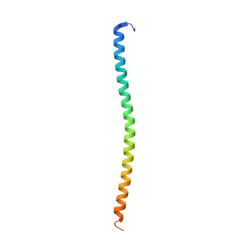
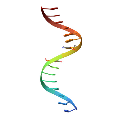
Structural basis for effects of CpA modifications on C/EBP beta binding of DNA.
Yang, J., Horton, J.R., Wang, D., Ren, R., Li, J., Sun, D., Huang, Y., Zhang, X., Blumenthal, R.M., Cheng, X.(2019) Nucleic Acids Res 47: 1774-1785
- PubMed: 30566668
- DOI: https://doi.org/10.1093/nar/gky1264
- Primary Citation of Related Structures:
6MG1, 6MG2, 6MG3 - PubMed Abstract:
CCAAT/enhancer binding proteins (C/EBPs) regulate gene expression in a variety of cells/tissues/organs, during a range of developmental stages, under both physiological and pathological conditions. C/EBP-related transcription factors have a consensus binding specificity of 5'-TTG-CG-CAA-3', with a central CpG/CpG and two outer CpA/TpG dinucleotides. Methylation of the CpG and CpA sites generates a DNA element with every pyrimidine having a methyl group in the 5-carbon position (thymine or 5-methylcytosine (5mC)). To understand the effects of both CpG and CpA modification on a centrally-important transcription factor, we show that C/EBPβ binds the methylated 8-bp element with modestly-increased (2.4-fold) binding affinity relative to the unmodified cognate sequence, while cytosine hydroxymethylation (particularly at the CpA sites) substantially decreased binding affinity (36-fold). The structure of C/EBPβ DNA binding domain in complex with methylated DNA revealed that the methyl groups of the 5mCpA/TpG make van der Waals contacts with Val285 in C/EBPβ. Arg289 recognizes the central 5mCpG by forming a methyl-Arg-G triad, and its conformation is constrained by Val285 and the 5mCpG methyl group. We substituted Val285 with Ala (V285A) in an Ala-Val dipeptide, to mimic the conserved Ala-Ala in many members of the basic leucine-zipper family of transcription factors, important in gene regulation, cell proliferation and oncogenesis. The V285A variant demonstrated a 90-fold binding preference for methylated DNA (particularly 5mCpA methylation) over the unmodified sequence. The smaller side chain of Ala285 permits Arg289 to adopt two alternative conformations, to interact in a similar fashion with either the central 5mCpG or the TpG of the opposite strand. Significantly, the best-studied cis-regulatory elements in RNA polymerase II promoters and enhancers have variable sequences corresponding to the central CpG or reduced to a single G:C base pair, but retain a conserved outer CpA sequence. Our analyses suggest an important modification-dependent CpA recognition by basic leucine-zipper transcription factors.
Organizational Affiliation:
Department of Molecular and Cellular Oncology, The University of Texas MD Anderson Cancer Center, Houston, TX 77030, USA.


