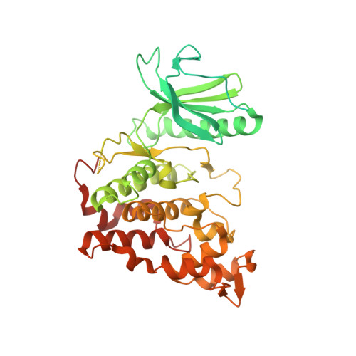
Structural Basis for the Selective Inhibition of Cdc2-Like Kinases by CX-4945.
Lee, J.Y., Yun, J.S., Kim, W.K., Chun, H.S., Jin, H., Cho, S., Chang, J.H.(2019) Biomed Res Int 2019: 6125068-6125068
- PubMed: 31531359
- DOI: https://doi.org/10.1155/2019/6125068
- Primary Citation of Related Structures:
6KHD, 6KHE, 6KHF - PubMed Abstract:
Cdc2-like kinases (CLKs) play a crucial role in the alternative splicing of eukaryotic pre-mRNAs through the phosphorylation of serine/arginine-rich proteins (SR proteins). Dysregulation of this processes is linked with various diseases including cancers, neurodegenerative diseases, and many genetic diseases. Thus, CLKs have been regarded to have a potential as a therapeutic target and significant efforts have been exerted to discover an effective inhibitor. In particular, the small molecule CX-4945, originally identified as an inhibitor of casein kinase 2 (CK2), was further revealed to have a strong CLK-inhibitory activity. Four isoforms of CLKs (CLK1, CLK2, CLK3, and CLK4) can be inhibited by CX-4945, with the highest inhibitory effect on CLK2. This study aimed to elucidate the structural basis of the selective inhibitory effect of CX-4945 on different isoforms of CLKs. We determined the crystal structures of CLK1, CLK2, and CLK3 in complex with CX-4945 at resolutions of 2.4 Å, 2.8 Å, and 2.6 Å, respectively. Comparative analysis revealed that CX-4945 was bound in the same active site pocket of the CLKs with similar interacting networks. Intriguingly, the active sites of CLK/CX-4945 complex structures had different sizes and electrostatic surface charge distributions. The active site of CLK1 was somewhat narrow and contained a negatively charged patch. CLK3 had a protruded Lys248 residue in the entrance of the active site pocket. In addition, Ala319, equivalent to Val324 (CLK1) and Val326 (CLK2), contributed to the weak hydrophobic interactions with the benzonaphthyridine ring of CX-4945. In contrast, the charge distribution pattern of CLK2 was the weakest, favoring its interactions with benzonaphthyridine ring. Thus, the relatively strong binding affinities of CX-4945 with CLK2 are consistent with its strong inhibitory effect defined in the previous study. These results may provide insights into structure-based drug discovery processes.
Organizational Affiliation:
Department of Biology Education, Kyungpook National University, 80 Daehak-ro, Buk-gu, Daegu 41566, Republic of Korea.


