6K08

Crystal structure of REV7(R124A/A135D) in complex with a Shieldin3 fragment


Experimental Data Snapshot

  • Method: X-RAY DIFFRACTION
  • Resolution: 2.31 Å
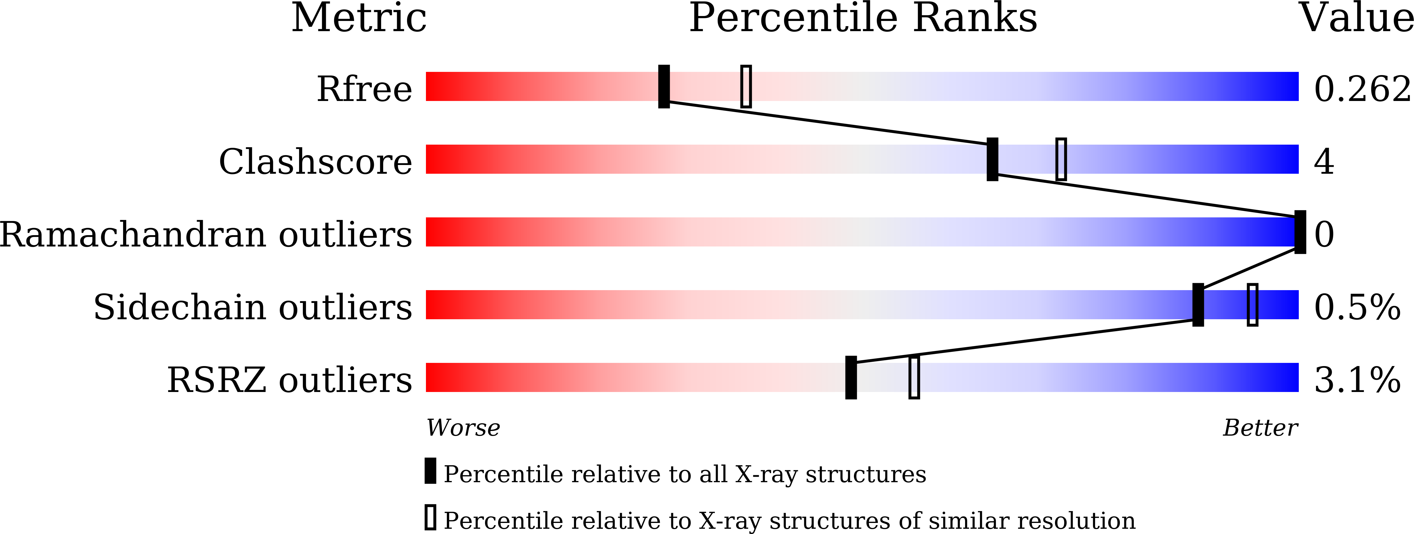
  • R-Value Free: 0.262 
  • R-Value Work: 0.210 
  • R-Value Observed: 0.213 

Starting Model: experimental
View more details

wwPDB Validation   3D Report Full Report


This is version 1.3 of the entry. See complete history


Literature

Structural basis for shieldin complex subunit 3-mediated recruitment of the checkpoint protein REV7 during DNA double-strand break repair.

Dai, Y.Zhang, F.Wang, L.Shan, S.Gong, Z.Zhou, Z.

(2020) J Biol Chem 295: 250-262

  • DOI: https://doi.org/10.1074/jbc.RA119.011464
  • Primary Citation of Related Structures:  
    6K07, 6K08

  • PubMed Abstract: 

    Shieldin complex subunit 3 (SHLD3) is the apical subunit of a recently-identified shieldin complex and plays a critical role in DNA double-strand break repair. To fulfill its function in DNA repair, SHLD3 interacts with the mitotic spindle assembly checkpoint protein REV7 homolog (REV7), but the details of this interaction remain obscure. Here, we present the crystal structures of REV7 in complex with SHLD3's REV7-binding domain (RBD) at 2.2-2.3 Å resolutions. The structures revealed that the ladle-shaped RBD in SHLD3 uses its N-terminal loop and C-terminal α-helix (αC-helix) in its interaction with REV7. The N-terminal loop exhibited a structure similar to those previously identified in other REV7-binding proteins, and the less-conserved αC-helix region adopted a distinct mode for binding REV7. In vitro and in vivo binding analyses revealed that the N-terminal loop and the αC-helix are both indispensable for high-affinity REV7 binding (with low-nanomolar affinity), underscoring the crucial role of SHLD3 αC-helix in protein binding. Moreover, binding kinetics analyses revealed that the REV7 "safety belt" region, which plays a role in binding other proteins, is essential for SHLD3-REV7 binding, as this region retards the dissociation of the RBD from the bound REV7. Together, the findings of our study reveal the molecular basis of the SHLD3-REV7 interaction and provide critical insights into how SHLD3 recognizes REV7.


  • Organizational Affiliation

    National Laboratory of Biomacromolecules, CAS Center for Excellence in Biomacromolecules, Institute of Biophysics, Chinese Academy of Sciences, Beijing 100101, China; Institute of Biophysics, University of Chinese Academy of Sciences, Beijing 100049, China.


Macromolecules
Find similar proteins by:  (by identity cutoff)  |  3D Structure
Entity ID: 1
MoleculeChains Sequence LengthOrganismDetailsImage
Mitotic spindle assembly checkpoint protein MAD2B219Homo sapiensMutation(s): 2 
Gene Names: MAD2L2MAD2BREV7
UniProt & NIH Common Fund Data Resources
Find proteins for Q9UI95 (Homo sapiens)
Explore Q9UI95 
Go to UniProtKB:  Q9UI95
PHAROS:  Q9UI95
GTEx:  ENSG00000116670 
Entity Groups  
Sequence Clusters30% Identity50% Identity70% Identity90% Identity95% Identity100% Identity
UniProt GroupQ9UI95
Sequence Annotations
Expand
  • Reference Sequence
Find similar proteins by:  (by identity cutoff)  |  3D Structure
Entity ID: 2
MoleculeChains Sequence LengthOrganismDetailsImage
Shieldin complex subunit 330Homo sapiensMutation(s): 0 
Gene Names: SHLD3FLJ26957RINN1
UniProt & NIH Common Fund Data Resources
Find proteins for Q6ZNX1 (Homo sapiens)
Explore Q6ZNX1 
Go to UniProtKB:  Q6ZNX1
PHAROS:  Q6ZNX1
GTEx:  ENSG00000253251 
Entity Groups  
Sequence Clusters30% Identity50% Identity70% Identity90% Identity95% Identity100% Identity
UniProt GroupQ6ZNX1
Sequence Annotations
Expand
  • Reference Sequence
Small Molecules
Ligands 1 Unique
IDChains Name / Formula / InChI Key2D Diagram3D Interactions
SO4
Query on SO4

Download Ideal Coordinates CCD File 
C [auth A]SULFATE ION
O4 S
QAOWNCQODCNURD-UHFFFAOYSA-L
Experimental Data & Validation

Experimental Data

  • Method: X-RAY DIFFRACTION
  • Resolution: 2.31 Å
  • R-Value Free: 0.262 
  • R-Value Work: 0.210 
  • R-Value Observed: 0.213 
  • Space Group: P 32 2 1
Unit Cell:
Length ( Å )Angle ( ˚ )
a = 60.64α = 90
b = 60.64β = 90
c = 133.48γ = 120
Software Package:
Software NamePurpose
PHENIXrefinement
XDSdata reduction
Aimlessdata scaling
PHASERphasing

Structure Validation

View Full Validation Report


Entry History & Funding Information

Deposition Data


Funding OrganizationLocationGrant Number
National Science Foundation (China)China--

Revision History  (Full details and data files)

  • Version 1.0: 2019-12-11
    Type: Initial release
  • Version 1.1: 2019-12-18
    Changes: Database references
  • Version 1.2: 2020-01-15
    Changes: Database references
  • Version 1.3: 2023-11-22
    Changes: Data collection, Database references, Refinement description