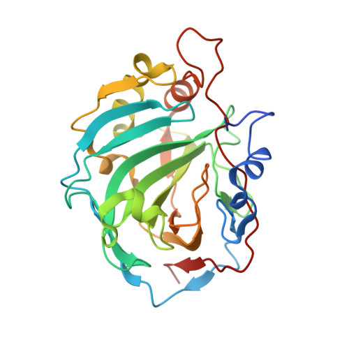
6HR3

Human Carbonic Anhydrase II in complex with 4-Propylbenzenesulfonamide


Experimental Data Snapshot

  • Method: X-RAY DIFFRACTION
  • Resolution: 1.02 Å
  • R-Value Free: 0.145 
  • R-Value Work: 0.129 
  • R-Value Observed: 0.130 

Starting Model: experimental
View more details

wwPDB Validation   3D Report Full Report


Ligand Structure Quality Assessment 


This is version 1.3 of the entry. See complete history


Literature

Conformational Changes in Alkyl Chains Determine the Thermodynamic and Kinetic Binding Profiles of Carbonic Anhydrase Inhibitors.

Glockner, S.Ngo, K.Sager, C.P.Hufner-Wulsdorf, T.Heine, A.Klebe, G.

(2020) ACS Chem Biol 15: 675-685

  • DOI: https://doi.org/10.1021/acschembio.9b00895
  • Primary Citation of Related Structures:  
    6GDC, 6GM9, 6HQX, 6HR3, 6HXD, 6I0W, 6I1U, 6I2F, 6I3E, 6SBH

  • PubMed Abstract: 

    Thermodynamics and kinetics of protein-ligand binding are both important aspects for the design of novel drug molecules. Presently, thermodynamic data are collected with isothermal titration calorimetry, while kinetic data are mostly derived from surface plasmon resonance. The new method of kinITC provides both thermodynamic and kinetic data from calorimetric titration measurements. The present study demonstrates the convenient collection of calorimetric data suitable for both thermodynamic and kinetic analysis for two series of congeneric ligands of human carbonic anhydrase II and correlates these findings with structural data obtained by macromolecular crystallography to shed light on the importance of shape complementarity for thermodynamics and kinetics governing a protein-ligand binding event. The study shows how minute chemical alterations change preferred ligand conformation and can be used to manipulate thermodynamic and kinetic signatures of binding. They give rise to the observation that analogous n -alkyl and n -alkyloxy derivatives of identical chain length swap their binding kinetic properties at unchanged binding affinity.


  • Organizational Affiliation

    Institut für Pharmazeutische Chemie, Philipps-Universität Marburg, Marbacher Weg 6, 35032 Marburg, Germany.


Macromolecules
Find similar proteins by:  (by identity cutoff)  |  3D Structure
Entity ID: 1
MoleculeChains Sequence LengthOrganismDetailsImage
Carbonic anhydrase 2265Homo sapiensMutation(s): 0 
Gene Names: CA2
EC: 4.2.1.1 (PDB Primary Data), 4.2.1.69 (UniProt)
UniProt & NIH Common Fund Data Resources
Find proteins for P00918 (Homo sapiens)
Explore P00918 
Go to UniProtKB:  P00918
PHAROS:  P00918
GTEx:  ENSG00000104267 
Entity Groups  
Sequence Clusters30% Identity50% Identity70% Identity90% Identity95% Identity100% Identity
UniProt GroupP00918
Sequence Annotations
Expand
  • Reference Sequence
Small Molecules
Binding Affinity Annotations 
IDSourceBinding Affinity
4JE BindingDB:  6HR3 Ki: 0.03 (nM) from 1 assay(s)
Kd: min: 5.78e+4, max: 6.06e+4 (nM) from 2 assay(s)
Experimental Data & Validation

Experimental Data

  • Method: X-RAY DIFFRACTION
  • Resolution: 1.02 Å
  • R-Value Free: 0.145 
  • R-Value Work: 0.129 
  • R-Value Observed: 0.130 
  • Space Group: P 1 21 1
Unit Cell:
Length ( Å )Angle ( ˚ )
a = 42.27α = 90
b = 41.519β = 104.59
c = 72.293γ = 90
Software Package:
Software NamePurpose
PHENIXrefinement
XDSdata reduction
XDSdata scaling
PHASERphasing
Cootmodel building

Structure Validation

View Full Validation Report


Ligand Structure Quality Assessment 


Entry History 

Deposition Data

Revision History  (Full details and data files)

  • Version 1.0: 2019-10-09
    Type: Initial release
  • Version 1.1: 2020-04-08
    Changes: Data collection, Database references
  • Version 1.2: 2020-07-29
    Type: Remediation
    Reason: Carbohydrate remediation
    Changes: Data collection, Derived calculations, Structure summary
  • Version 1.3: 2024-01-24
    Changes: Data collection, Database references, Refinement description, Structure summary