6HHG

Crystal Structure of AKT1 in Complex with Covalent-Allosteric AKT Inhibitor 27


Experimental Data Snapshot

  • Method: X-RAY DIFFRACTION
  • Resolution: 2.30 Å
  • R-Value Free: 0.246 
  • R-Value Work: 0.210 
  • R-Value Observed: 0.212 

Starting Model: experimental
View more details

wwPDB Validation   3D Report Full Report


Ligand Structure Quality Assessment 


This is version 1.2 of the entry. See complete history


Literature

Structural and chemical insights into the covalent-allosteric inhibition of the protein kinase Akt.

Uhlenbrock, N.Smith, S.Weisner, J.Landel, I.Lindemann, M.Le, T.A.Hardick, J.Gontla, R.Scheinpflug, R.Czodrowski, P.Janning, P.Depta, L.Quambusch, L.Muller, M.P.Engels, B.Rauh, D.

(2019) Chem Sci 10: 3573-3585

  • DOI: https://doi.org/10.1039/c8sc05212c
  • Primary Citation of Related Structures:  
    6HHG, 6HHH, 6HHI, 6HHJ

  • PubMed Abstract: 

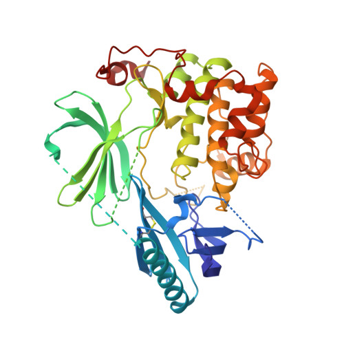
    The Ser/Thr kinase Akt (Protein Kinase B/PKB) is a master switch in cellular signal transduction pathways. Its downstream signaling influences cell proliferation, cell growth, and apoptosis, rendering Akt a prominent drug target. The unique activation mechanism of Akt involves a change of the relative orientation of its N-terminal pleckstrin homology (PH) and the kinase domain and makes this kinase suitable for highly specific allosteric modulation. Here we present a unique set of crystal structures of covalent-allosteric interdomain inhibitors in complex with full-length Akt and report the structure-based design, synthesis, biological and pharmacological evaluation of a focused library of these innovative inhibitors.


  • Organizational Affiliation

    Faculty of Chemistry and Chemical Biology , TU Dortmund University , Drug Discovery Hub Dortmund (DDHD) am Zentrum für integrierte Wirkstoffforschung (ZIW) , Otto-Hahn-Strasse 4a , 44227 Dortmund , Germany . Email: daniel.rauh@tu-dortmund.de ; http://www.ddhdortmund.de ; www.twitter.com/DDHDortmund.


Macromolecules
Find similar proteins by:  (by identity cutoff)  |  3D Structure
Entity ID: 1
MoleculeChains Sequence LengthOrganismDetailsImage
RAC-alpha serine/threonine-protein kinase446Homo sapiensMutation(s): 3 
Gene Names: AKT1PKBRAC
EC: 2.7.11.1
UniProt & NIH Common Fund Data Resources
Find proteins for P31749 (Homo sapiens)
Explore P31749 
Go to UniProtKB:  P31749
GTEx:  ENSG00000142208 
Entity Groups  
Sequence Clusters30% Identity50% Identity70% Identity90% Identity95% Identity100% Identity
UniProt GroupP31749
Sequence Annotations
Expand
  • Reference Sequence
Small Molecules
Ligands 1 Unique
IDChains Name / Formula / InChI Key2D Diagram3D Interactions
G4T
Query on G4T

Download Ideal Coordinates CCD File 
B [auth A]~{N}-[2-chloranyl-5-[[1-[[4-(5-oxidanylidene-3-phenyl-6~{H}-1,6-naphthyridin-2-yl)phenyl]methyl]piperidin-4-yl]carbamoylamino]phenyl]propanamide
C36 H35 Cl N6 O3
JIITZXKZXZZBIZ-UHFFFAOYSA-N
Experimental Data & Validation

Experimental Data

  • Method: X-RAY DIFFRACTION
  • Resolution: 2.30 Å
  • R-Value Free: 0.246 
  • R-Value Work: 0.210 
  • R-Value Observed: 0.212 
  • Space Group: P 21 21 21
Unit Cell:
Length ( Å )Angle ( ˚ )
a = 70.82α = 90
b = 71.23β = 90
c = 91.24γ = 90
Software Package:
Software NamePurpose
PHENIXrefinement
XDSdata reduction
XSCALEdata scaling
PHASERphasing

Structure Validation

View Full Validation Report


Ligand Structure Quality Assessment 


Entry History & Funding Information

Deposition Data


Funding OrganizationLocationGrant Number
German Federal Ministry for Education and ResearchGermanyBMBF 01GS08104
German Federal Ministry for Education and ResearchGermany01ZX1303C

Revision History  (Full details and data files)

  • Version 1.0: 2019-02-20
    Type: Initial release
  • Version 1.1: 2019-05-01
    Changes: Data collection, Database references
  • Version 1.2: 2024-01-17
    Changes: Data collection, Database references, Refinement description