6BN7

Crystal structure of DDB1-CRBN-BRD4(BD1) complex bound to dBET23 PROTAC.


Experimental Data Snapshot

  • Method: X-RAY DIFFRACTION
  • Resolution: 3.50 Å
  • R-Value Free: 0.256 
  • R-Value Work: 0.212 
  • R-Value Observed: 0.214 

Starting Models: experimental
View more details

wwPDB Validation   3D Report Full Report


Ligand Structure Quality Assessment 


This is version 1.5 of the entry. See complete history


Literature

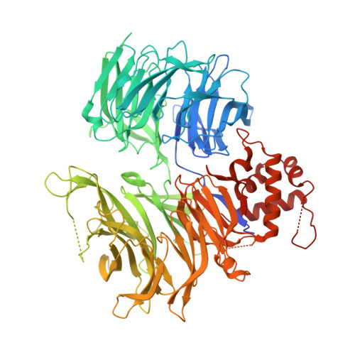
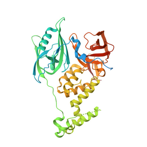
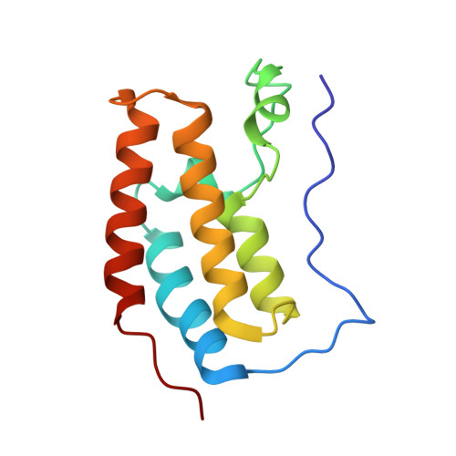
Plasticity in binding confers selectivity in ligand-induced protein degradation.

Nowak, R.P.DeAngelo, S.L.Buckley, D.He, Z.Donovan, K.A.An, J.Safaee, N.Jedrychowski, M.P.Ponthier, C.M.Ishoey, M.Zhang, T.Mancias, J.D.Gray, N.S.Bradner, J.E.Fischer, E.S.

(2018) Nat Chem Biol 14: 706-714

  • DOI: https://doi.org/10.1038/s41589-018-0055-y
  • Primary Citation of Related Structures:  
    6BN7, 6BN8, 6BN9, 6BNB, 6BOY

  • PubMed Abstract: 

    Heterobifunctional small-molecule degraders that induce protein degradation through ligase-mediated ubiquitination have shown considerable promise as a new pharmacological modality. However, we currently lack a detailed understanding of the molecular basis for target recruitment and selectivity, which is critically required to enable rational design of degraders. Here we utilize a comprehensive characterization of the ligand-dependent CRBN-BRD4 interaction to demonstrate that binding between proteins that have not evolved to interact is plastic. Multiple X-ray crystal structures show that plasticity results in several distinct low-energy binding conformations that are selectively bound by ligands. We demonstrate that computational protein-protein docking can reveal the underlying interprotein contacts and inform the design of a BRD4 selective degrader that can discriminate between highly homologous BET bromodomains. Our findings that plastic interprotein contacts confer selectivity for ligand-induced protein dimerization provide a conceptual framework for the development of heterobifunctional ligands.


  • Organizational Affiliation

    Department of Cancer Biology, Dana-Farber Cancer Institute, Boston, MA, USA.


Macromolecules
Find similar proteins by:  (by identity cutoff)  |  3D Structure
Entity ID: 1
MoleculeChains Sequence LengthOrganismDetailsImage
DNA damage-binding protein 1864Homo sapiensMutation(s): 0 
Gene Names: DDB1XAP1
UniProt & NIH Common Fund Data Resources
Find proteins for Q16531 (Homo sapiens)
Explore Q16531 
Go to UniProtKB:  Q16531
GTEx:  ENSG00000167986 
Entity Groups  
Sequence Clusters30% Identity50% Identity70% Identity90% Identity95% Identity100% Identity
UniProt GroupQ16531
Sequence Annotations
Expand
  • Reference Sequence
Find similar proteins by:  (by identity cutoff)  |  3D Structure
Entity ID: 2
MoleculeChains Sequence LengthOrganismDetailsImage
Protein cereblon463Homo sapiensMutation(s): 0 
Gene Names: CRBNAD-006
UniProt & NIH Common Fund Data Resources
Find proteins for Q96SW2 (Homo sapiens)
Explore Q96SW2 
Go to UniProtKB:  Q96SW2
GTEx:  ENSG00000113851 
Entity Groups  
Sequence Clusters30% Identity50% Identity70% Identity90% Identity95% Identity100% Identity
UniProt GroupQ96SW2
Sequence Annotations
Expand
  • Reference Sequence
Find similar proteins by:  (by identity cutoff)  |  3D Structure
Entity ID: 3
MoleculeChains Sequence LengthOrganismDetailsImage
Bromodomain-containing protein 4127Homo sapiensMutation(s): 1 
Gene Names: BRD4HUNK1
UniProt & NIH Common Fund Data Resources
Find proteins for O60885 (Homo sapiens)
Explore O60885 
Go to UniProtKB:  O60885
GTEx:  ENSG00000141867 
Entity Groups  
Sequence Clusters30% Identity50% Identity70% Identity90% Identity95% Identity100% Identity
UniProt GroupO60885
Sequence Annotations
Expand
  • Reference Sequence
Small Molecules
Ligands 2 Unique
IDChains Name / Formula / InChI Key2D Diagram3D Interactions
RN3 (Subject of Investigation/LOI)
Query on RN3

Download Ideal Coordinates CCD File 
E [auth B]methyl {(6S)-4-(4-chlorophenyl)-2-[(8-{[({2-[(3S)-2,6-dioxopiperidin-3-yl]-1,3-dioxo-2,3-dihydro-1H-isoindol-4-yl}oxy)acetyl]amino}octyl)carbamoyl]-3,9-dimethyl-6H-thieno[3,2-f][1,2,4]triazolo[4,3-a][1,4]diazepin-6-yl}acetate
C43 H45 Cl N8 O9 S
ZIHIUFZFPMILIG-VMPREFPWSA-N
ZN
Query on ZN

Download Ideal Coordinates CCD File 
D [auth B]ZINC ION
Zn
PTFCDOFLOPIGGS-UHFFFAOYSA-N
Experimental Data & Validation

Experimental Data

  • Method: X-RAY DIFFRACTION
  • Resolution: 3.50 Å
  • R-Value Free: 0.256 
  • R-Value Work: 0.212 
  • R-Value Observed: 0.214 
  • Space Group: P 65 2 2
Unit Cell:
Length ( Å )Angle ( ˚ )
a = 115.571α = 90
b = 115.571β = 90
c = 596.315γ = 120
Software Package:
Software NamePurpose
PHENIXrefinement
XDSdata reduction
Aimlessdata scaling
PHASERphasing

Structure Validation

View Full Validation Report


Ligand Structure Quality Assessment 


Entry History & Funding Information

Deposition Data


Funding OrganizationLocationGrant Number
National Institutes of Health/National Cancer Institute (NIH/NCI)United StatesNCI R01CA214608
The Harvard University William F. Milton FundUnited States--
the Friends of Dana FarberUnited States--
Claudia Adams Barr Program for Innovative Cancer ResearchUnited States--
Linde Family FoundationUnited States--

Revision History  (Full details and data files)

  • Version 1.0: 2018-05-30
    Type: Initial release
  • Version 1.1: 2018-08-01
    Changes: Data collection, Database references
  • Version 1.2: 2018-11-07
    Changes: Data collection, Database references
  • Version 1.3: 2019-02-20
    Changes: Author supporting evidence, Data collection
  • Version 1.4: 2019-12-04
    Changes: Author supporting evidence
  • Version 1.5: 2023-10-04
    Changes: Data collection, Database references, Refinement description