Hydration Structures of the Human Protein Kinase CK2 alpha Clarified by Joint Neutron and X-ray Crystallography.
Shibazaki, C., Arai, S., Shimizu, R., Saeki, M., Kinoshita, T., Ostermann, A., Schrader, T.E., Kurosaki, Y., Sunami, T., Kuroki, R., Adachi, M.(2018) J Mol Biol 430: 5094-5104
- PubMed: 30359582
- DOI: https://doi.org/10.1016/j.jmb.2018.09.018
- Primary Citation of Related Structures:
5ZN0, 5ZN1, 5ZN2, 5ZN3, 5ZN4, 5ZN5 - PubMed Abstract:
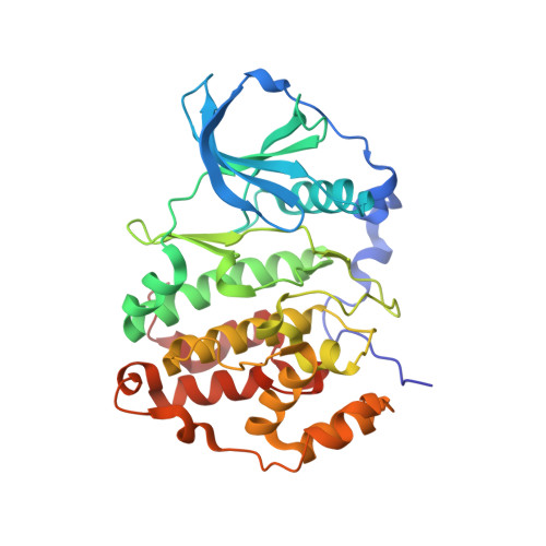
Casein kinase 2 (CK2) has broad phosphorylation activity against various regulatory proteins, which are important survival factors in eukaryotic cells. To clarify the hydration structure and catalytic mechanism of CK2, we determined the crystal structure of the alpha subunit of human CK2 containing hydrogen and deuterium atoms using joint neutron (1.9 Å resolution) and X-ray (1.1 Å resolution) crystallography. The analysis revealed the structure of conserved water molecules at the active site and a long potential hydrogen bonding network originating from the catalytic Asp156 that is well known to enhance the nucleophilicity of the substrate OH group to the γ-phospho group of ATP by proton elimination. His148 and Asp214 conserved in the protein kinase family are located in the middle of the network. The water molecule forming a hydrogen bond with Asp214 appears to be deformed. In addition, mutational analysis of His148 in CK2 showed significant reductions by 40%-75% in the catalytic efficiency with similar affinity for ATP. Likewise, remarkable reductions to less than 5% were shown by corresponding mutations on His131 in death-associated protein kinase 1, which belongs to a group different from that of CK2. These findings shed new light on the catalytic mechanism of protein kinases in which the hydrogen bond network through the C-terminal domain may assist the general base catalyst to extract a proton with a link to the bulk solvent via intermediates of a pair of residues.
Organizational Affiliation:
National Institutes for Quantum and Radiological Science and Technology, 2-4 Shirakata, Tokai, Ibaraki 319-1106, Japan.


