5Z2T

Crystal structure of DNA-bound DUX4-HD2


Experimental Data Snapshot

  • Method: X-RAY DIFFRACTION
  • Resolution: 2.62 Å
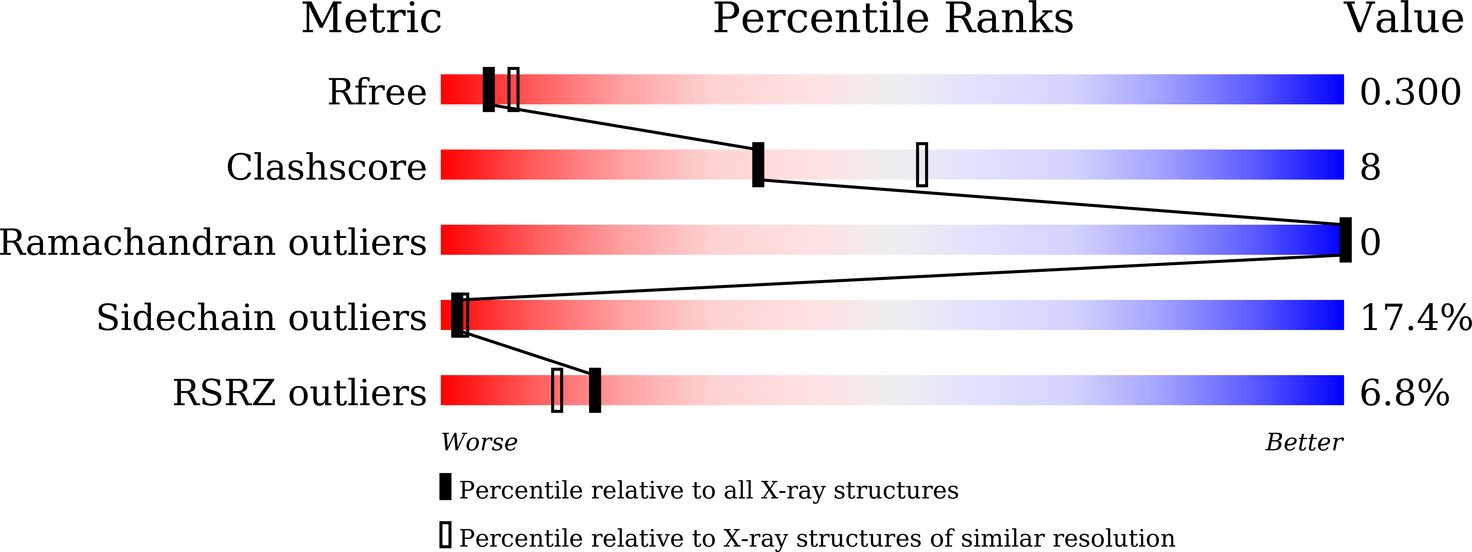
  • R-Value Free: 0.299 
  • R-Value Work: 0.276 
  • R-Value Observed: 0.278 

Starting Model: experimental
View more details

wwPDB Validation   3D Report Full Report


This is version 1.2 of the entry. See complete history


Literature

Structural basis of DUX4/IGH-driven transactivation.

Dong, X.Zhang, W.Wu, H.Huang, J.Zhang, M.Wang, P.Zhang, H.Chen, Z.Chen, S.J.Meng, G.

(2018) Leukemia 32: 1466-1476

  • DOI: https://doi.org/10.1038/s41375-018-0093-1
  • Primary Citation of Related Structures:  
    5Z2S, 5Z2T

  • PubMed Abstract: 

    Oncogenic fusions are major drivers in leukemogenesis and may serve as potent targets for treatment. DUX4/IGHs have been shown to trigger the abnormal expression of ERG alt through binding to DUX4-Responsive-Element (DRE), which leads to B-cell differentiation arrest and a full-fledged B-ALL. Here, we determined the crystal structures of Apo- and DNA DRE -bound DUX4 HD2 and revealed a clamp-like transactivation mechanism via the double homeobox domain. Biophysical characterization showed that mutations in the interacting interfaces significantly impaired the DNA binding affinity of DUX4 homeobox. These mutations, when introduced into DUX4/IGH, abrogated its transactivation activity in Reh cells. More importantly, the structure-based mutants significantly impaired the inhibitory effects of DUX4/IGH upon B-cell differentiation in mouse progenitor cells. All these results help to define a key DUX4/IGH-DRE recognition/step in B-ALL.


  • Organizational Affiliation

    State Key Laboratory of Medical Genomics, Shanghai Institute of Hematology, Rui-Jin Hospital, Shanghai JiaoTong University School of Medicine and School of Life Sciences and Biotechnology, Shanghai JiaoTong University, 197 Ruijin Er Road, Shanghai, 200025, China.


Macromolecules

Find similar proteins by:  (by identity cutoff)  |  3D Structure
Entity ID: 1
MoleculeChains Sequence LengthOrganismDetailsImage
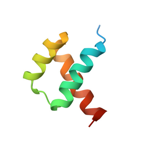
Double homeobox protein 4A [auth C],
D
64Homo sapiensMutation(s): 0 
Gene Names: DUX4DUX10
UniProt & NIH Common Fund Data Resources
Find proteins for Q9UBX2 (Homo sapiens)
Explore Q9UBX2 
Go to UniProtKB:  Q9UBX2
PHAROS:  Q9UBX2
GTEx:  ENSG00000260596 
Entity Groups  
Sequence Clusters30% Identity50% Identity70% Identity90% Identity95% Identity100% Identity
UniProt GroupQ9UBX2
Sequence Annotations
Expand
  • Reference Sequence

Find similar nucleic acids by:  Sequence   |   3D Structure  

Entity ID: 2
MoleculeChains LengthOrganismImage
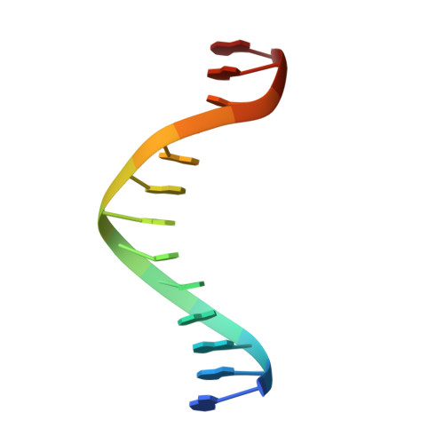
5'-D(*TP*TP*CP*TP*AP*AP*TP*CP*TP*AP*AP*TP*CP*TP*T)-3'B [auth E]15synthetic construct
Sequence Annotations
Expand
  • Reference Sequence

Find similar nucleic acids by:  Sequence   |   3D Structure  

Entity ID: 3
MoleculeChains LengthOrganismImage
5'-D(P*AP*AP*GP*AP*TP*TP*AP*GP*AP*TP*TP*AP*GP*T)-3'C [auth F]14synthetic construct
Sequence Annotations
Expand
  • Reference Sequence
Experimental Data & Validation

Experimental Data

  • Method: X-RAY DIFFRACTION
  • Resolution: 2.62 Å
  • R-Value Free: 0.299 
  • R-Value Work: 0.276 
  • R-Value Observed: 0.278 
  • Space Group: P 41 21 2
Unit Cell:
Length ( Å )Angle ( ˚ )
a = 51.61α = 90
b = 51.61β = 90
c = 166.55γ = 90
Software Package:
Software NamePurpose
PHENIXrefinement
MOSFLMdata reduction
SCALAdata scaling
PHASERphasing

Structure Validation

View Full Validation Report


Entry History & Funding Information

Deposition Data


Funding OrganizationLocationGrant Number
National Natural Science Foundation of ChinaChina81770142
National Natural Science Foundation of ChinaChina81370620
National Natural Science Foundation of ChinaChina81570120

Revision History  (Full details and data files)

  • Version 1.0: 2018-04-04
    Type: Initial release
  • Version 1.1: 2018-06-20
    Changes: Data collection, Database references
  • Version 1.2: 2023-11-22
    Changes: Data collection, Database references, Refinement description