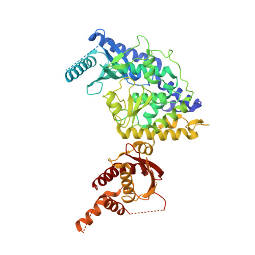
5WU2

Crystal structure of human Tut1 bound with BaUTP, form I


Experimental Data Snapshot

  • Method: X-RAY DIFFRACTION
  • Resolution: 2.95 Å
  • R-Value Free: 0.294 
  • R-Value Work: 0.246 
  • R-Value Observed: 0.248 

wwPDB Validation   3D Report Full Report


Ligand Structure Quality Assessment 


This is version 1.2 of the entry. See complete history


Literature

Crystal structures of U6 snRNA-specific terminal uridylyltransferase

Yamashita, S.Takagi, Y.Nagaike, T.Tomita, K.

(2017) Nat Commun 8: 15788-15788

  • DOI: https://doi.org/10.1038/ncomms15788
  • Primary Citation of Related Structures:  
    5WU1, 5WU2, 5WU3, 5WU4, 5WU5, 5WU6

  • PubMed Abstract: 

    The terminal uridylyltransferase, TUT1, builds or repairs the 3'-oligo-uridylylated tail of U6 snRNA. The 3'-oligo-uridylylated tail is the Lsm-binding site for U4/U6 di-snRNP formation and U6 snRNA recycling for pre-mRNA splicing. Here, we report crystallographic and biochemical analyses of human TUT1, which revealed the mechanisms for the specific uridylylation of the 3'-end of U6 snRNA by TUT1. The O 2 and O 4 atoms of the UTP base form hydrogen bonds with the conserved His and Asn in the catalytic pocket, respectively, and TUT1 preferentially incorporates UMP onto the 3'-end of RNAs. TUT1 recognizes the entire U6 snRNA molecule by its catalytic domains, N-terminal RNA-recognition motifs and a previously unidentified C-terminal RNA-binding domain. Each domain recognizes specific regions within U6 snRNA, and the recognition is coupled with the domain movements and U6 snRNA structural changes. Hence, TUT1 functions as the U6 snRNA-specific terminal uridylyltransferase required for pre-mRNA splicing.


  • Organizational Affiliation

    Department of Computational Biology and Medical Sciences, Graduate School of Frontier Sciences, the University of Tokyo, Kashiwa, Chiba 277-8562, Japan.


Macromolecules
Find similar proteins by:  (by identity cutoff)  |  3D Structure
Entity ID: 1
MoleculeChains Sequence LengthOrganismDetailsImage
Speckle targeted PIP5K1A-regulated poly(A) polymerase
A, B
573Homo sapiensMutation(s): 4 
Gene Names: TUT1RBM21
EC: 2.7.7.19 (PDB Primary Data), 2.7.7.52 (PDB Primary Data)
UniProt & NIH Common Fund Data Resources
Find proteins for Q9H6E5 (Homo sapiens)
Explore Q9H6E5 
Go to UniProtKB:  Q9H6E5
PHAROS:  Q9H6E5
GTEx:  ENSG00000149016 
Entity Groups  
Sequence Clusters30% Identity50% Identity70% Identity90% Identity95% Identity100% Identity
UniProt GroupQ9H6E5
Sequence Annotations
Expand
  • Reference Sequence
Experimental Data & Validation

Experimental Data

  • Method: X-RAY DIFFRACTION
  • Resolution: 2.95 Å
  • R-Value Free: 0.294 
  • R-Value Work: 0.246 
  • R-Value Observed: 0.248 
  • Space Group: P 21 21 21
Unit Cell:
Length ( Å )Angle ( ˚ )
a = 77.33α = 90
b = 88.4β = 90
c = 184.19γ = 90
Software Package:
Software NamePurpose
PHENIXrefinement
XSCALEdata scaling
PDB_EXTRACTdata extraction
XDSdata reduction
SOLVEphasing

Structure Validation

View Full Validation Report


Ligand Structure Quality Assessment 


Entry History & Funding Information

Deposition Data


Funding OrganizationLocationGrant Number
JSPSJapan26251009

Revision History  (Full details and data files)

  • Version 1.0: 2017-05-31
    Type: Initial release
  • Version 1.1: 2017-06-21
    Changes: Database references
  • Version 1.2: 2024-03-20
    Changes: Data collection, Database references, Derived calculations