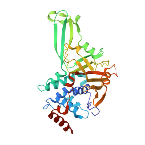
Structure-Guided Development of a Potent and Selective Non-covalent Active-Site Inhibitor of USP7.
Lamberto, I., Liu, X., Seo, H.S., Schauer, N.J., Iacob, R.E., Hu, W., Das, D., Mikhailova, T., Weisberg, E.L., Engen, J.R., Anderson, K.C., Chauhan, D., Dhe-Paganon, S., Buhrlage, S.J.(2017) Cell Chem Biol 24: 1490-1500.e11
- PubMed: 29056421
- DOI: https://doi.org/10.1016/j.chembiol.2017.09.003
- Primary Citation of Related Structures:
5VS6, 5VSB, 5VSK - PubMed Abstract:
Deubiquitinating enzymes (DUBs) have garnered significant attention as drug targets in the last 5-10 years. The excitement stems in large part from the powerful ability of DUB inhibitors to promote degradation of oncogenic proteins, especially proteins that are challenging to directly target but which are stabilized by DUB family members. Highly optimized and well-characterized DUB inhibitors have thus become highly sought after tools. Most reported DUB inhibitors, however, are polypharmacological agents possessing weak (micromolar) potency toward their primary target, limiting their utility in target validation and mechanism studies. Due to a lack of high-resolution DUB⋅small-molecule ligand complex structures, no structure-guided optimization efforts have been reported for a mammalian DUB. Here, we report a small-molecule⋅ubiquitin-specific protease (USP) family DUB co-structure and rapid design of potent and selective inhibitors of USP7 guided by the structure. Interestingly, the compounds are non-covalent active-site inhibitors.
Organizational Affiliation:
Department of Cancer Biology, Dana-Farber Cancer Institute, Boston, MA 02215, USA.


