5VP7

Crystal structure of human KRAS G12A mutant in complex with GDP


Experimental Data Snapshot

  • Method: X-RAY DIFFRACTION
  • Resolution: 1.70 Å
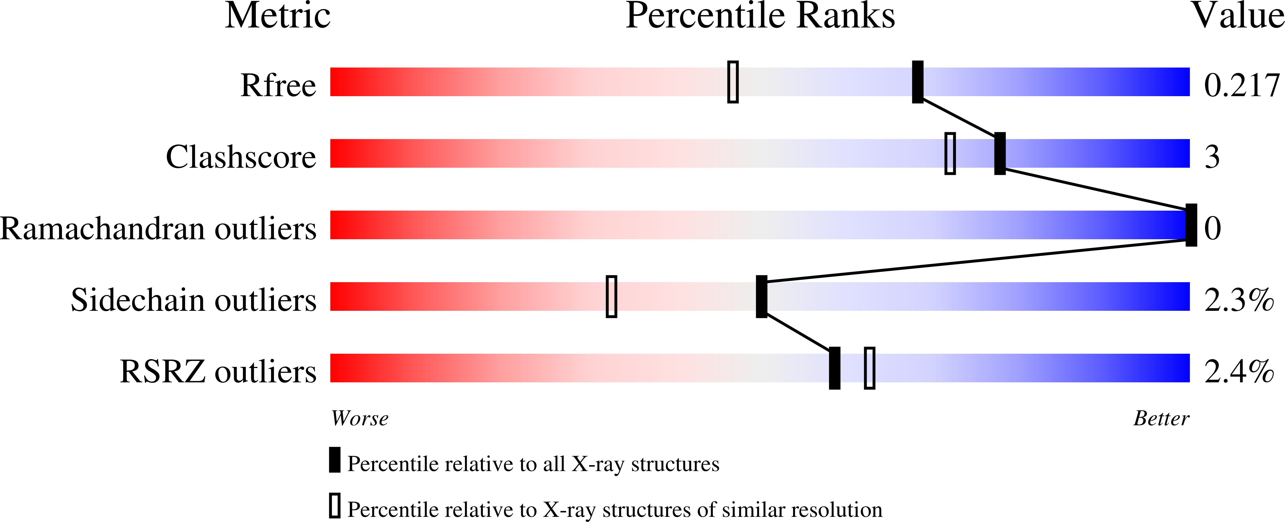
  • R-Value Free: 0.207 
  • R-Value Work: 0.171 
  • R-Value Observed: 0.173 

Starting Model: experimental
View more details

wwPDB Validation   3D Report Full Report


Ligand Structure Quality Assessment 


This is version 1.2 of the entry. See complete history


Literature

Structural insight into the rearrangement of the switch I region in GTP-bound G12A K-Ras.

Xu, S.Long, B.N.Boris, G.H.Chen, A.Ni, S.Kennedy, M.A.

(2017) Acta Crystallogr D Struct Biol 73: 970-984

  • DOI: https://doi.org/10.1107/S2059798317015418
  • Primary Citation of Related Structures:  
    5VP7, 5VPI, 5VPY, 5VPZ, 5VQ0, 5VQ1, 5VQ2, 5VQ6, 5VQ8, 5W22

  • PubMed Abstract: 

    K-Ras, a molecular switch that regulates cell growth, apoptosis and metabolism, is activated when it undergoes a conformation change upon binding GTP and is deactivated following the hydrolysis of GTP to GDP. Hydrolysis of GTP in water is accelerated by coordination to K-Ras, where GTP adopts a high-energy conformation approaching the transition state. The G12A mutation reduces intrinsic K-Ras GTP hydrolysis by an unexplained mechanism. Here, crystal structures of G12A K-Ras in complex with GDP, GTP, GTPγS and GppNHp, and of Q61A K-Ras in complex with GDP, are reported. In the G12A K-Ras-GTP complex, the switch I region undergoes a significant reorganization such that the Tyr32 side chain points towards the GTP-binding pocket and forms a hydrogen bond to the GTP γ-phosphate, effectively stabilizing GTP in its precatalytic state, increasing the activation energy required to reach the transition state and contributing to the reduced intrinsic GTPase activity of G12A K-Ras mutants.


  • Organizational Affiliation

    Department of Chemistry and Biochemistry, Miami University, Oxford, OH 45056, USA.


Macromolecules
Find similar proteins by:  (by identity cutoff)  |  3D Structure
Entity ID: 1
MoleculeChains Sequence LengthOrganismDetailsImage
GTPase KRasA,
B [auth F]
170Homo sapiensMutation(s): 2 
Gene Names: KRASKRAS2RASK2
UniProt & NIH Common Fund Data Resources
Find proteins for P01116 (Homo sapiens)
Explore P01116 
Go to UniProtKB:  P01116
GTEx:  ENSG00000133703 
Entity Groups  
Sequence Clusters30% Identity50% Identity70% Identity90% Identity95% Identity100% Identity
UniProt GroupP01116
Sequence Annotations
Expand
  • Reference Sequence
Small Molecules
Ligands 4 Unique
IDChains Name / Formula / InChI Key2D Diagram3D Interactions
GDP
Query on GDP

Download Ideal Coordinates CCD File 
C [auth A],
G [auth F]
GUANOSINE-5'-DIPHOSPHATE
C10 H15 N5 O11 P2
QGWNDRXFNXRZMB-UUOKFMHZSA-N
PGE
Query on PGE

Download Ideal Coordinates CCD File 
E [auth A]TRIETHYLENE GLYCOL
C6 H14 O4
ZIBGPFATKBEMQZ-UHFFFAOYSA-N
MG
Query on MG

Download Ideal Coordinates CCD File 
D [auth A],
H [auth F]
MAGNESIUM ION
Mg
JLVVSXFLKOJNIY-UHFFFAOYSA-N
NA
Query on NA

Download Ideal Coordinates CCD File 
F [auth A],
I [auth F]
SODIUM ION
Na
FKNQFGJONOIPTF-UHFFFAOYSA-N
Experimental Data & Validation

Experimental Data

  • Method: X-RAY DIFFRACTION
  • Resolution: 1.70 Å
  • R-Value Free: 0.207 
  • R-Value Work: 0.171 
  • R-Value Observed: 0.173 
  • Space Group: H 3
Unit Cell:
Length ( Å )Angle ( ˚ )
a = 93.773α = 90
b = 93.773β = 90
c = 119.918γ = 120
Software Package:
Software NamePurpose
REFMACrefinement
SCALAdata scaling
PDB_EXTRACTdata extraction
iMOSFLMdata reduction
PHASERphasing

Structure Validation

View Full Validation Report


Ligand Structure Quality Assessment 


Entry History 

Deposition Data

Revision History  (Full details and data files)

  • Version 1.0: 2017-12-06
    Type: Initial release
  • Version 1.1: 2017-12-20
    Changes: Database references
  • Version 1.2: 2023-10-04
    Changes: Data collection, Database references, Derived calculations, Refinement description