5UNJ

Structure of Human Liver Receptor Homolog 1 in complex with PGC1a and RJW100

  • Classification: NUCLEAR PROTEIN
  • Organism(s): Homo sapiens
  • Expression System: Escherichia coli
  • Mutation(s): No 

  • Deposited: 2017-01-31 Released: 2017-04-19 
  • Deposition Author(s): Mays, S.G., Ortlund, E.A.
  • Funding Organization(s): National Institutes of Health/National Institute of Diabetes and Digestive and Kidney Disease (NIH/NIDDK), National Institutes of Health/National Institute of General Medical Sciences (NIH/NIGMS)

Experimental Data Snapshot

  • Method: X-RAY DIFFRACTION
  • Resolution: 1.96 Å
  • R-Value Free: 0.227 
  • R-Value Work: 0.199 
  • R-Value Observed: 0.201 

Starting Model: experimental
View more details

wwPDB Validation   3D Report Full Report


Ligand Structure Quality Assessment 


This is version 1.4 of the entry. See complete history


Literature

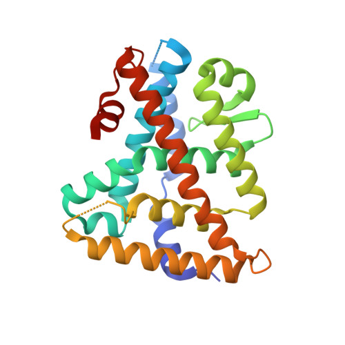
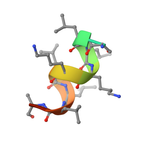
Structure and Dynamics of the Liver Receptor Homolog 1-PGC1 alpha Complex.

Mays, S.G.Okafor, C.D.Tuntland, M.L.Whitby, R.J.Dharmarajan, V.Stec, J.Griffin, P.R.Ortlund, E.A.

(2017) Mol Pharmacol 92: 1-11

  • DOI: https://doi.org/10.1124/mol.117.108514
  • Primary Citation of Related Structures:  
    5UNJ

  • PubMed Abstract: 

    Peroxisome proliferator-activated gamma coactivator 1- α (PGC1 α ) regulates energy metabolism by directly interacting with transcription factors to modulate gene expression. Among the PGC1 α binding partners is liver receptor homolog 1 (LRH-1; NR5A2), an orphan nuclear hormone receptor that controls lipid and glucose homeostasis. Although PGC1 α is known to bind and activate LRH-1, mechanisms through which PGC1 α changes LRH-1 conformation to drive transcription are unknown. Here, we used biochemical and structural methods to interrogate the LRH-1-PGC1 α complex. Purified, full-length LRH-1, as well as isolated ligand binding domain, bound to PGC1 α with higher affinity than to the coactivator, nuclear receptor coactivator-2 (Tif2), in coregulator peptide recruitment assays. We present the first crystal structure of the LRH-1-PGC1 α complex, which depicts several hydrophobic contacts and a strong charge clamp at the interface between these partners. In molecular dynamics simulations, PGC1 α induced correlated atomic motion throughout the entire LRH-1 activation function surface, which was dependent on charge-clamp formation. In contrast, Tif2 induced weaker signaling at the activation function surface than PGC1 α but promoted allosteric signaling from the helix 6/ β -sheet region of LRH-1 to the activation function surface. These studies are the first to probe mechanisms underlying the LRH-1-PGC1 α interaction and may illuminate strategies for selective therapeutic targeting of PGC1 α -dependent LRH-1 signaling pathways.


  • Organizational Affiliation

    Department of Biochemistry, Emory University School of Medicine, Atlanta, Georgia (S.G.M., C.D.O., M.L.T., E.A.O.); School of Chemistry, University of Southampton, Southampton, United Kingdom (R.J.W., J.S.); and Department of Molecular Medicine, Scripps Research Institute, Jupiter, Florida (V.D., P.R.G.).


Macromolecules
Find similar proteins by:  (by identity cutoff)  |  3D Structure
Entity ID: 1
MoleculeChains Sequence LengthOrganismDetailsImage
Nuclear receptor subfamily 5 group A member 2245Homo sapiensMutation(s): 0 
Gene Names: NR5A2B1FCPFFTF
UniProt & NIH Common Fund Data Resources
Find proteins for O00482 (Homo sapiens)
Explore O00482 
Go to UniProtKB:  O00482
GTEx:  ENSG00000116833 
Entity Groups  
Sequence Clusters30% Identity50% Identity70% Identity90% Identity95% Identity100% Identity
UniProt GroupO00482
Sequence Annotations
Expand
  • Reference Sequence

Find similar proteins by:  Sequence   |   3D Structure  

Entity ID: 2
MoleculeChains Sequence LengthOrganismDetailsImage
Peroxisome proliferator-activated gamma coactivator 1-alphaB [auth C]14Homo sapiensMutation(s): 0 
UniProt & NIH Common Fund Data Resources
Find proteins for Q9UBK2 (Homo sapiens)
Explore Q9UBK2 
Go to UniProtKB:  Q9UBK2
GTEx:  ENSG00000109819 
Entity Groups  
Sequence Clusters30% Identity50% Identity70% Identity90% Identity95% Identity100% Identity
UniProt GroupQ9UBK2
Sequence Annotations
Expand
  • Reference Sequence
Small Molecules
Ligands 1 Unique
IDChains Name / Formula / InChI Key2D Diagram3D Interactions
RJW
Query on RJW

Download Ideal Coordinates CCD File 
C [auth A](1R,3aR,6aR)-5-hexyl-4-phenyl-3a-(1-phenylethenyl)-1,2,3,3a,6,6a-hexahydropentalen-1-ol
C28 H34 O
ZFXMYHPLTQTTFW-REUBFRLUSA-N
Binding Affinity Annotations 
IDSourceBinding Affinity
RJW BindingDB:  5UNJ Ki: 374 (nM) from 1 assay(s)
EC50: min: 251.19, max: 1500 (nM) from 4 assay(s)
Experimental Data & Validation

Experimental Data

  • Method: X-RAY DIFFRACTION
  • Resolution: 1.96 Å
  • R-Value Free: 0.227 
  • R-Value Work: 0.199 
  • R-Value Observed: 0.201 
  • Space Group: P 21 21 2
Unit Cell:
Length ( Å )Angle ( ˚ )
a = 66.18α = 90
b = 84.027β = 90
c = 45.355γ = 90
Software Package:
Software NamePurpose
HKL-2000data scaling
PHENIXrefinement
PDB_EXTRACTdata extraction
PHENIXphasing
DENZOdata reduction
SCALEPACKdata scaling

Structure Validation

View Full Validation Report


Ligand Structure Quality Assessment 


Entry History & Funding Information

Deposition Data


Funding OrganizationLocationGrant Number
National Institutes of Health/National Institute of Diabetes and Digestive and Kidney Disease (NIH/NIDDK)United StatesR01DK095750
National Institutes of Health/National Institute of Diabetes and Digestive and Kidney Disease (NIH/NIDDK)United StatesF31DK111171
National Institutes of Health/National Institute of General Medical Sciences (NIH/NIGMS)United StatesT32GM008602
National Institutes of Health/National Institute of General Medical Sciences (NIH/NIGMS)United StatesNIH K12GM000680

Revision History  (Full details and data files)

  • Version 1.0: 2017-04-19
    Type: Initial release
  • Version 1.1: 2017-06-07
    Changes: Database references
  • Version 1.2: 2017-09-20
    Changes: Author supporting evidence, Refinement description
  • Version 1.3: 2019-12-25
    Changes: Author supporting evidence
  • Version 1.4: 2023-10-04
    Changes: Data collection, Database references, Refinement description