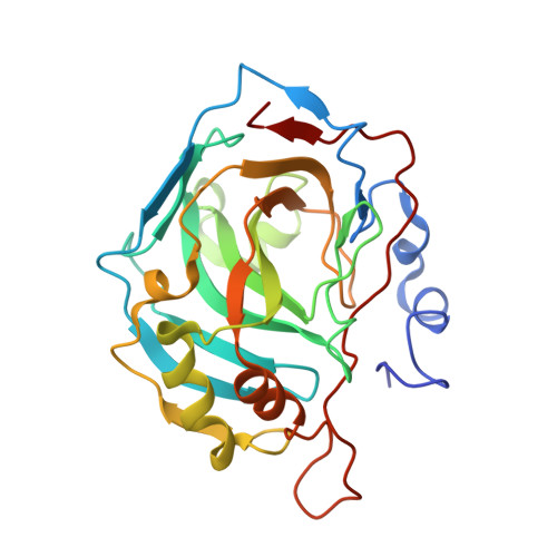
5THJ

Crystal Structure of 2-hydroxycyclohepta-2,4,6-trien-1-one bound to human carbonic anhydrase 2


Experimental Data Snapshot

  • Method: X-RAY DIFFRACTION
  • Resolution: 1.50 Å
  • R-Value Free: 0.189 
  • R-Value Work: 0.154 
  • R-Value Observed: 0.156 

Starting Model: experimental
View more details

wwPDB Validation   3D Report Full Report


Ligand Structure Quality Assessment 


This is version 1.4 of the entry. See complete history


Literature

Effect of donor atom identity on metal-binding pharmacophore coordination.

Dick, B.L.Patel, A.McCammon, J.A.Cohen, S.M.

(2017) J Biol Inorg Chem 22: 605-613

  • DOI: https://doi.org/10.1007/s00775-017-1454-3
  • Primary Citation of Related Structures:  
    5TH4, 5THI, 5THJ, 5THN, 5TI0

  • PubMed Abstract: 

    The inhibition and binding of three metal-binding pharmacophores (MBPs), 2-hydroxycyclohepta-2,4,6-trien-1-one (tropolone), 2-mercaptopyridine-N-oxide (1,2-HOPTO), and 2-hydroxycyclohepta-2,4,6-triene-1-thione (thiotropolone) to human carbonic anhydrase II (hCAII) and a mutant protein hCAII L198G were investigated. These MBPs displayed bidentate coordination to the active site Zn(II) metal ion, but the MBPs respond to the mutation of L198G differently, as characterized by inhibition activity assays and X-ray crystallography. The L198G mutation increases the active site volume thereby decreasing the steric pressure exerted on MBPs upon binding, allowing changes in MBP coordination to be observed. When comparing the binding mode of tropolone to thiotropolone or 1,2-HOPTO (O,O versus O,S donor sets), structural modifications of the hCAII active site were shown to have a stronger effect on MBPs with an O,O versus O,S donor set. These findings were corroborated with density functional theory (DFT) calculations of model coordination complexes. These results suggest that the MBP binding geometry is a malleable interaction, particularly for certain ligands, and that the identity of the donor atoms influences the response of the ligand to changes in the protein active site environment. Understanding underlying interactions between a MBP and a metalloenzyme active site may aid in the design and development of potent metalloenzyme inhibitors.


  • Organizational Affiliation

    Department of Chemistry and Biochemistry, University of California San Diego, 9500 Gilman Drive, La Jolla, CA, 92093-0358, USA.


Macromolecules
Find similar proteins by:  (by identity cutoff)  |  3D Structure
Entity ID: 1
MoleculeChains Sequence LengthOrganismDetailsImage
Carbonic anhydrase 2260Homo sapiensMutation(s): 0 
Gene Names: CA2
EC: 4.2.1.1 (PDB Primary Data), 4.2.1.69 (UniProt)
UniProt & NIH Common Fund Data Resources
Find proteins for P00918 (Homo sapiens)
Explore P00918 
Go to UniProtKB:  P00918
PHAROS:  P00918
GTEx:  ENSG00000104267 
Entity Groups  
Sequence Clusters30% Identity50% Identity70% Identity90% Identity95% Identity100% Identity
UniProt GroupP00918
Sequence Annotations
Expand
  • Reference Sequence
Small Molecules
Ligands 6 Unique
IDChains Name / Formula / InChI Key2D Diagram3D Interactions
MBO
Query on MBO

Download Ideal Coordinates CCD File 
D [auth A]MERCURIBENZOIC ACID
C7 H5 Hg O2
FVFZSVRSDNUCGG-UHFFFAOYSA-N
CIT
Query on CIT

Download Ideal Coordinates CCD File 
L [auth A]CITRIC ACID
C6 H8 O7
KRKNYBCHXYNGOX-UHFFFAOYSA-N
0TR
Query on 0TR

Download Ideal Coordinates CCD File 
C [auth A]2-HYDROXYCYCLOHEPTA-2,4,6-TRIEN-1-ONE
C7 H6 O2
MDYOLVRUBBJPFM-UHFFFAOYSA-N
GOL
Query on GOL

Download Ideal Coordinates CCD File 
K [auth A]GLYCEROL
C3 H8 O3
PEDCQBHIVMGVHV-UHFFFAOYSA-N
DMS
Query on DMS

Download Ideal Coordinates CCD File 
E [auth A]
F [auth A]
G [auth A]
H [auth A]
I [auth A]
E [auth A],
F [auth A],
G [auth A],
H [auth A],
I [auth A],
J [auth A]
DIMETHYL SULFOXIDE
C2 H6 O S
IAZDPXIOMUYVGZ-UHFFFAOYSA-N
ZN
Query on ZN

Download Ideal Coordinates CCD File 
B [auth A]ZINC ION
Zn
PTFCDOFLOPIGGS-UHFFFAOYSA-N
Experimental Data & Validation

Experimental Data

  • Method: X-RAY DIFFRACTION
  • Resolution: 1.50 Å
  • R-Value Free: 0.189 
  • R-Value Work: 0.154 
  • R-Value Observed: 0.156 
  • Space Group: P 1 21 1
Unit Cell:
Length ( Å )Angle ( ˚ )
a = 42.283α = 90
b = 41.34β = 104
c = 71.848γ = 90
Software Package:
Software NamePurpose
REFMACrefinement
APEXdata collection
PHASERphasing

Structure Validation

View Full Validation Report


Ligand Structure Quality Assessment 


Entry History & Funding Information

Deposition Data


Funding OrganizationLocationGrant Number
National Institutes of Health/National Institute of General Medical Sciences (NIH/NIGMS)United StatesR01 GM098435

Revision History  (Full details and data files)

  • Version 1.0: 2017-04-26
    Type: Initial release
  • Version 1.1: 2017-06-07
    Changes: Database references
  • Version 1.2: 2017-09-20
    Changes: Author supporting evidence
  • Version 1.3: 2019-12-25
    Changes: Author supporting evidence
  • Version 1.4: 2023-10-04
    Changes: Data collection, Database references, Refinement description