5ODT

Aurora-A in complex with TACC3


Experimental Data Snapshot

  • Method: X-RAY DIFFRACTION
  • Resolution: 2.02 Å
  • R-Value Free: 0.211 
  • R-Value Work: 0.178 
  • R-Value Observed: 0.179 

Starting Model: experimental
View more details

wwPDB Validation   3D Report Full Report


Ligand Structure Quality Assessment 


This is version 1.3 of the entry. See complete history


Literature

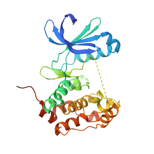
Mitotic spindle association of TACC3 requires Aurora-A-dependent stabilization of a cryptic alpha-helix.

Burgess, S.G.Mukherjee, M.Sabir, S.Joseph, N.Gutierrez-Caballero, C.Richards, M.W.Huguenin-Dezot, N.Chin, J.W.Kennedy, E.J.Pfuhl, M.Royle, S.J.Gergely, F.Bayliss, R.

(2018) EMBO J 37

  • DOI: https://doi.org/10.15252/embj.201797902
  • Primary Citation of Related Structures:  
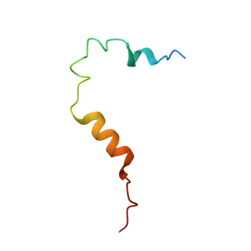
    5ODT

  • PubMed Abstract: 

    Aurora-A regulates the recruitment of TACC3 to the mitotic spindle through a phospho-dependent interaction with clathrin heavy chain (CHC). Here, we describe the structural basis of these interactions, mediated by three motifs in a disordered region of TACC3. A hydrophobic docking motif binds to a previously uncharacterized pocket on Aurora-A that is blocked in most kinases. Abrogation of the docking motif causes a delay in late mitosis, consistent with the cellular distribution of Aurora-A complexes. Phosphorylation of Ser558 engages a conformational switch in a second motif from a disordered state, needed to bind the kinase active site, into a helical conformation. The helix extends into a third, adjacent motif that is recognized by a helical-repeat region of CHC, not a recognized phospho-reader domain. This potentially widespread mechanism of phospho-recognition provides greater flexibility to tune the molecular details of the interaction than canonical recognition motifs that are dominated by phosphate binding.


  • Organizational Affiliation

    Astbury Centre for Structural Molecular Biology, School of Molecular and Cellular Biology, Faculty of Biological Sciences, University of Leeds, Leeds, UK.


Macromolecules
Find similar proteins by:  (by identity cutoff)  |  3D Structure
Entity ID: 1
MoleculeChains Sequence LengthOrganismDetailsImage
Aurora kinase A283Homo sapiensMutation(s): 3 
Gene Names: AURKAAIKAIRK1ARK1AURAAYK1BTAKIAK1STK15STK6
EC: 2.7.11.1
UniProt & NIH Common Fund Data Resources
Find proteins for O14965 (Homo sapiens)
Explore O14965 
Go to UniProtKB:  O14965
GTEx:  ENSG00000087586 
Entity Groups  
Sequence Clusters30% Identity50% Identity70% Identity90% Identity95% Identity100% Identity
UniProt GroupO14965
Sequence Annotations
Expand
  • Reference Sequence
Find similar proteins by:  (by identity cutoff)  |  3D Structure
Entity ID: 2
MoleculeChains Sequence LengthOrganismDetailsImage
Transforming acidic coiled-coil-containing protein 346Homo sapiensMutation(s): 0 
Gene Names: TACC3ERIC1
UniProt & NIH Common Fund Data Resources
Find proteins for Q9Y6A5 (Homo sapiens)
Explore Q9Y6A5 
Go to UniProtKB:  Q9Y6A5
GTEx:  ENSG00000013810 
Entity Groups  
Sequence Clusters30% Identity50% Identity70% Identity90% Identity95% Identity100% Identity
UniProt GroupQ9Y6A5
Sequence Annotations
Expand
  • Reference Sequence
Small Molecules
Ligands 3 Unique
IDChains Name / Formula / InChI Key2D Diagram3D Interactions
ADP
Query on ADP

Download Ideal Coordinates CCD File 
C [auth A]ADENOSINE-5'-DIPHOSPHATE
C10 H15 N5 O10 P2
XTWYTFMLZFPYCI-KQYNXXCUSA-N
SO4
Query on SO4

Download Ideal Coordinates CCD File 
G [auth A],
H [auth A],
I [auth A],
J [auth B]
SULFATE ION
O4 S
QAOWNCQODCNURD-UHFFFAOYSA-L
GOL
Query on GOL

Download Ideal Coordinates CCD File 
D [auth A],
E [auth A],
F [auth A]
GLYCEROL
C3 H8 O3
PEDCQBHIVMGVHV-UHFFFAOYSA-N
Experimental Data & Validation

Experimental Data

  • Method: X-RAY DIFFRACTION
  • Resolution: 2.02 Å
  • R-Value Free: 0.211 
  • R-Value Work: 0.178 
  • R-Value Observed: 0.179 
  • Space Group: I 2 3
Unit Cell:
Length ( Å )Angle ( ˚ )
a = 137.04α = 90
b = 137.04β = 90
c = 137.04γ = 90
Software Package:
Software NamePurpose
PHENIXrefinement
xia2data reduction
xia2data scaling
PHASERphasing

Structure Validation

View Full Validation Report


Ligand Structure Quality Assessment 


Entry History & Funding Information

Deposition Data


Funding OrganizationLocationGrant Number
Cancer Research UKUnited KingdomC24461/A12772

Revision History  (Full details and data files)

  • Version 1.0: 2018-03-14
    Type: Initial release
  • Version 1.1: 2018-03-21
    Changes: Database references
  • Version 1.2: 2018-04-25
    Changes: Data collection, Database references
  • Version 1.3: 2024-01-17
    Changes: Data collection, Database references, Refinement description