5M8P

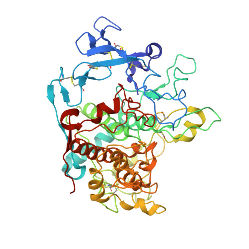
Crystal structure of human tyrosinase related protein 1 in complex with tyrosine


Experimental Data Snapshot

  • Method: X-RAY DIFFRACTION
  • Resolution: 2.80 Å
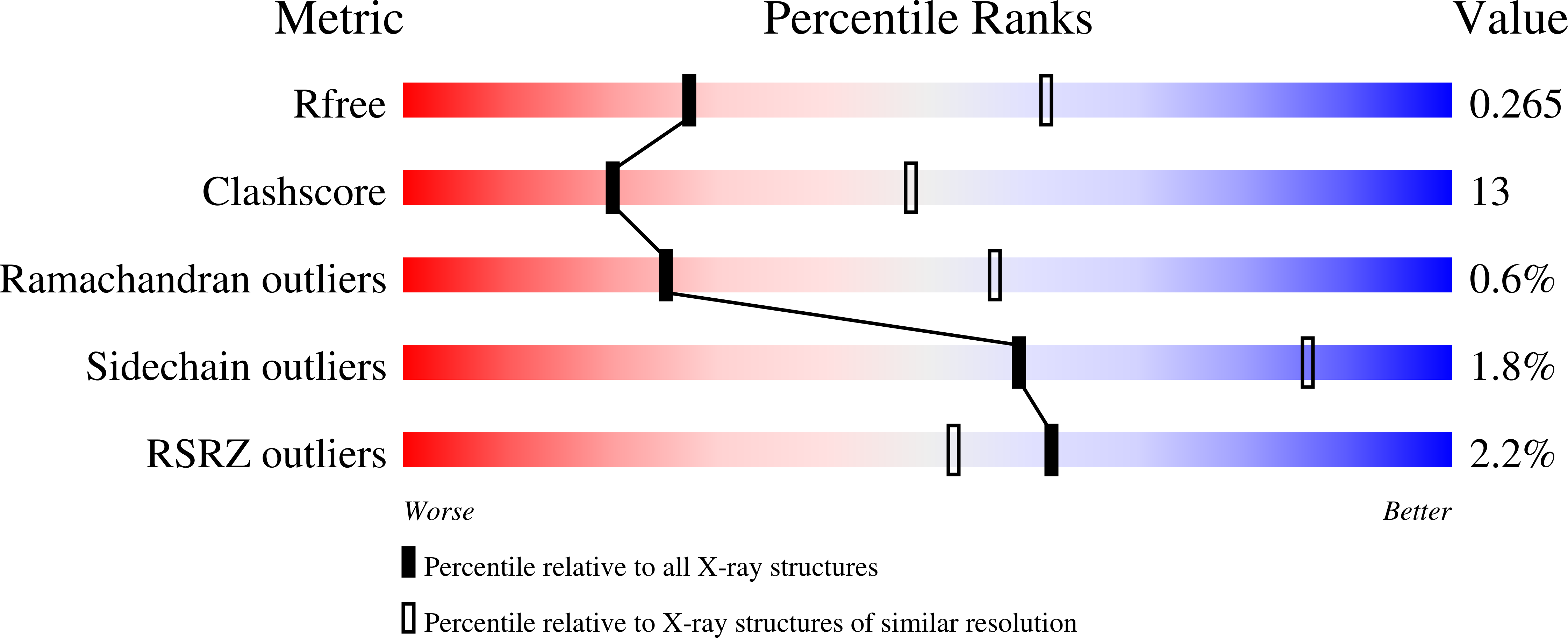
  • R-Value Free: 0.262 
  • R-Value Work: 0.205 
  • R-Value Observed: 0.207 

wwPDB Validation   3D Report Full Report


Ligand Structure Quality Assessment 


This is version 4.0 of the entry. See complete history


Literature

Structure of Human Tyrosinase Related Protein 1 Reveals a Binuclear Zinc Active Site Important for Melanogenesis.

Lai, X.Wichers, H.J.Soler-Lopez, M.Dijkstra, B.W.

(2017) Angew Chem Int Ed Engl 56: 9812-9815

  • DOI: https://doi.org/10.1002/anie.201704616
  • Primary Citation of Related Structures:  
    5M8L, 5M8M, 5M8N, 5M8O, 5M8P, 5M8Q, 5M8R, 5M8T

  • PubMed Abstract: 

    Tyrosinase-related protein 1 (TYRP1) is one of three tyrosinase-like glycoenzymes in human melanocytes that are key to the production of melanin, the compound responsible for the pigmentation of skin, eye, and hair. Difficulties with producing these enzymes in pure form have hampered the understanding of their activity and the effect of mutations that cause albinism and pigmentation disorders. Herein we show that the typical tyrosinase-like subdomain of TYRP1 contains two zinc ions in the active site instead of copper ions as found in tyrosinases, which explains why TYRP1 does not exhibit tyrosinase redox activity. In addition, the structures reveal for the first time that the Cys-rich subdomain, which is unique to vertebrate melanogenic proteins, has an epidermal growth factor-like fold and is tightly associated with the tyrosinase subdomain. Our structures suggest that most albinism-related mutations of TYRP1 affect its stability or activity.


  • Organizational Affiliation

    Laboratory of Biophysical Chemistry, University of Groningen, Nijenborgh 7, 9747 AG, Groningen, The Netherlands.


Macromolecules
Find similar proteins by:  (by identity cutoff)  |  3D Structure
Entity ID: 1
MoleculeChains Sequence LengthOrganismDetailsImage
5,6-dihydroxyindole-2-carboxylic acid oxidase
A, B, C, D
446Homo sapiensMutation(s): 0 
Gene Names: TYRP1CAS2TYRPTYRRP
EC: 1.14.18
UniProt & NIH Common Fund Data Resources
Find proteins for P17643 (Homo sapiens)
Explore P17643 
Go to UniProtKB:  P17643
PHAROS:  P17643
GTEx:  ENSG00000107165 
Entity Groups  
Sequence Clusters30% Identity50% Identity70% Identity90% Identity95% Identity100% Identity
UniProt GroupP17643
Glycosylation
Glycosylation Sites: 6Go to GlyGen: P17643-1
Sequence Annotations
Expand
  • Reference Sequence
Oligosaccharides

Help

Entity ID: 2
MoleculeChains Length2D Diagram Glycosylation3D Interactions
2-acetamido-2-deoxy-beta-D-glucopyranose-(1-4)-[alpha-L-fucopyranose-(1-6)]2-acetamido-2-deoxy-beta-D-glucopyranose
E, J, O, U
3N-Glycosylation
Glycosylation Resources
GlyTouCan:  G21290RB
GlyCosmos:  G21290RB
GlyGen:  G21290RB
Entity ID: 3
MoleculeChains Length2D Diagram Glycosylation3D Interactions
alpha-L-fucopyranose-(1-6)-2-acetamido-2-deoxy-beta-D-glucopyranose
F, K, Q
2N-Glycosylation
Glycosylation Resources
GlyTouCan:  G86851RC
GlyCosmos:  G86851RC
GlyGen:  G86851RC
Entity ID: 4
MoleculeChains Length2D Diagram Glycosylation3D Interactions
2-acetamido-2-deoxy-beta-D-glucopyranose-(1-4)-2-acetamido-2-deoxy-beta-D-glucopyranose
G, H, I, N, P
G, H, I, N, P, R, W, Y
2N-Glycosylation
Glycosylation Resources
GlyTouCan:  G42666HT
GlyCosmos:  G42666HT
GlyGen:  G42666HT
Entity ID: 5
MoleculeChains Length2D Diagram Glycosylation3D Interactions
alpha-D-mannopyranose-(1-4)-2-acetamido-2-deoxy-beta-D-glucopyranose-(1-4)-2-acetamido-2-deoxy-beta-D-glucopyranose
L
3N-Glycosylation
Glycosylation Resources
GlyTouCan:  G62182OO
GlyCosmos:  G62182OO
GlyGen:  G62182OO
Entity ID: 6
MoleculeChains Length2D Diagram Glycosylation3D Interactions
alpha-D-mannopyranose-(1-3)-[alpha-D-mannopyranose-(1-6)]alpha-D-mannopyranose-(1-4)-2-acetamido-2-deoxy-beta-D-glucopyranose-(1-4)-2-acetamido-2-deoxy-beta-D-glucopyranose
M, X
5N-Glycosylation
Glycosylation Resources
GlyTouCan:  G21381MC
GlyCosmos:  G21381MC
GlyGen:  G21381MC
Entity ID: 7
MoleculeChains Length2D Diagram Glycosylation3D Interactions
2-acetamido-2-deoxy-beta-D-glucopyranose-(1-3)-2-acetamido-2-deoxy-beta-D-glucopyranose
S
2N/A
Glycosylation Resources
GlyTouCan:  G79924RM
GlyCosmos:  G79924RM
GlyGen:  G79924RM
Entity ID: 8
MoleculeChains Length2D Diagram Glycosylation3D Interactions
alpha-D-mannopyranose-(1-3)-2-acetamido-2-deoxy-beta-D-glucopyranose-(1-4)-2-acetamido-2-deoxy-beta-D-glucopyranose
T
3N-Glycosylation
Glycosylation Resources
GlyTouCan:  G66019PC
GlyCosmos:  G66019PC
GlyGen:  G66019PC
Entity ID: 9
MoleculeChains Length2D Diagram Glycosylation3D Interactions
alpha-D-mannopyranose-(1-4)-2-acetamido-2-deoxy-beta-D-glucopyranose-(1-4)-[alpha-L-fucopyranose-(5-6)]2-acetamido-2-deoxy-beta-D-glucopyranose
V
4N/AN-Glycosylation
Small Molecules
Ligands 4 Unique
IDChains Name / Formula / InChI Key2D Diagram3D Interactions
NAG
Query on NAG

Download Ideal Coordinates CCD File 
Z [auth A]2-acetamido-2-deoxy-beta-D-glucopyranose
C8 H15 N O6
OVRNDRQMDRJTHS-FMDGEEDCSA-N
TYR
Query on TYR

Download Ideal Coordinates CCD File 
AA [auth A],
EA [auth B],
IA [auth C],
LA [auth D]
TYROSINE
C9 H11 N O3
OUYCCCASQSFEME-QMMMGPOBSA-N
MAN
Query on MAN

Download Ideal Coordinates CCD File 
HA [auth C]alpha-D-mannopyranose
C6 H12 O6
WQZGKKKJIJFFOK-PQMKYFCFSA-N
ZN
Query on ZN

Download Ideal Coordinates CCD File 
BA [auth A]
CA [auth A]
DA [auth A]
FA [auth B]
GA [auth B]
BA [auth A],
CA [auth A],
DA [auth A],
FA [auth B],
GA [auth B],
JA [auth C],
KA [auth C],
MA [auth D],
NA [auth D]
ZINC ION
Zn
PTFCDOFLOPIGGS-UHFFFAOYSA-N
Experimental Data & Validation

Experimental Data

  • Method: X-RAY DIFFRACTION
  • Resolution: 2.80 Å
  • R-Value Free: 0.262 
  • R-Value Work: 0.205 
  • R-Value Observed: 0.207 
  • Space Group: P 21 21 21
Unit Cell:
Length ( Å )Angle ( ˚ )
a = 89.516α = 90
b = 140.322β = 90
c = 191.676γ = 90
Software Package:
Software NamePurpose
REFMACrefinement
PDB_EXTRACTdata extraction
PHENIXrefinement
SCALAdata scaling
PHENIXphasing

Structure Validation

View Full Validation Report


Ligand Structure Quality Assessment 


Entry History 

Deposition Data

Revision History  (Full details and data files)

  • Version 1.0: 2017-07-12
    Type: Initial release
  • Version 1.1: 2017-08-09
    Changes: Database references
  • Version 2.0: 2017-11-15
    Changes: Advisory, Atomic model, Derived calculations, Non-polymer description, Structure summary
  • Version 2.1: 2019-10-16
    Changes: Data collection
  • Version 3.0: 2020-07-29
    Type: Remediation
    Reason: Carbohydrate remediation
    Changes: Advisory, Atomic model, Data collection, Derived calculations, Structure summary
  • Version 4.0: 2022-12-07
    Changes: Advisory, Atomic model, Data collection, Database references, Derived calculations